• 文献检索
  • 文档翻译
  • 深度研究
  • 学术资讯
  • Suppr Zotero 插件Zotero 插件
  • 邀请有礼
  • 套餐&价格
  • 历史记录
应用&插件
Suppr Zotero 插件Zotero 插件浏览器插件Mac 客户端Windows 客户端微信小程序
定价
高级版会员购买积分包购买API积分包
服务
文献检索文档翻译深度研究API 文档MCP 服务
关于我们
关于 Suppr公司介绍联系我们用户协议隐私条款
关注我们

Suppr 超能文献

核心技术专利:CN118964589B侵权必究
粤ICP备2023148730 号-1Suppr @ 2026

文献检索

告别复杂PubMed语法,用中文像聊天一样搜索,搜遍4000万医学文献。AI智能推荐,让科研检索更轻松。

立即免费搜索

文件翻译

保留排版,准确专业,支持PDF/Word/PPT等文件格式,支持 12+语言互译。

免费翻译文档

深度研究

AI帮你快速写综述,25分钟生成高质量综述,智能提取关键信息,辅助科研写作。

立即免费体验

可植入肌源性生物材料和工程化肌肉组织功能评估中的成像方法

Imaging Approaches in Functional Assessment of Implantable Myogenic Biomaterials and Engineered Muscle Tissue.

作者信息

Edmunds Kyle J, Gargiulo Paolo

机构信息

Institute for Biomedical and Neural Engineering, University of Reykjavík.

Institute for Biomedical and Neural Engineering, University of Reykjavík; University Hospital Landspítali, Reykjavík, Iceland.

出版信息

Eur J Transl Myol. 2015 Mar 11;25(2):4847. doi: 10.4081/ejtm.2015.4847.

DOI:10.4081/ejtm.2015.4847
PMID:26913149
原文链接:https://pmc.ncbi.nlm.nih.gov/articles/PMC4749010/
Abstract

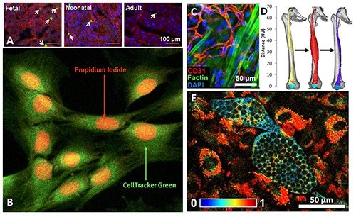
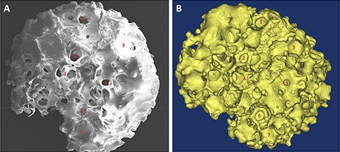
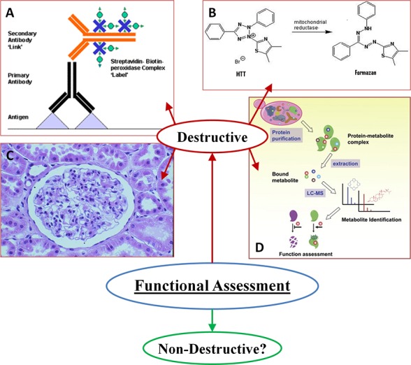
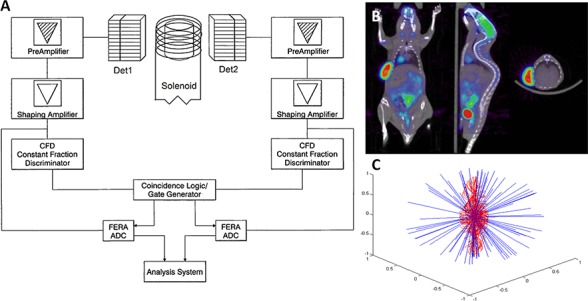
The fields of tissue engineering and regenerative medicine utilize implantable biomaterials and engineered tissues to regenerate damaged cells or replace lost tissues. There are distinct challenges in all facets of this research, but functional assessments and monitoring of such complex environments as muscle tissues present the current strategic priority. Many extant methods for addressing these questions result in the destruction or alteration of tissues or cell populations under investigation. Modern advances in non-invasive imaging modalities present opportunities to rethink some of the anachronistic methods, however, their standard employment may not be optimal when considering advancements in myology. New image analysis protocols and/or combinations of established modalities need to be addressed. This review focuses on efficacies and limitations of available imaging modalities to the functional assessment of implantable myogenic biomaterials and engineered muscle tissues.

摘要

组织工程和再生医学领域利用可植入生物材料和工程组织来再生受损细胞或替代缺失组织。这项研究的各个方面都存在独特的挑战,但对肌肉组织等复杂环境进行功能评估和监测是当前的战略重点。许多解决这些问题的现有方法会导致所研究的组织或细胞群体被破坏或改变。然而,非侵入性成像方式的现代进展为重新思考一些过时的方法提供了机会,不过,在考虑肌肉学的进展时,其标准应用可能并非最佳。需要探讨新的图像分析方案和/或现有方式的组合。本综述重点关注现有成像方式在可植入肌源性生物材料和工程肌肉组织功能评估方面的功效和局限性。

https://cdn.ncbi.nlm.nih.gov/pmc/blobs/f53c/4749010/e822e9527873/ejtm-2015-2-4847-g004.jpg
https://cdn.ncbi.nlm.nih.gov/pmc/blobs/f53c/4749010/2c08a3d3b4d8/ejtm-2015-2-4847-g001.jpg
https://cdn.ncbi.nlm.nih.gov/pmc/blobs/f53c/4749010/1d603ea5e9ce/ejtm-2015-2-4847-g002.jpg
https://cdn.ncbi.nlm.nih.gov/pmc/blobs/f53c/4749010/fb9cbe67bab2/ejtm-2015-2-4847-g003.jpg
https://cdn.ncbi.nlm.nih.gov/pmc/blobs/f53c/4749010/e822e9527873/ejtm-2015-2-4847-g004.jpg

相似文献

1
Imaging Approaches in Functional Assessment of Implantable Myogenic Biomaterials and Engineered Muscle Tissue.可植入肌源性生物材料和工程化肌肉组织功能评估中的成像方法
Eur J Transl Myol. 2015 Mar 11;25(2):4847. doi: 10.4081/ejtm.2015.4847.
2
Imaging challenges in biomaterials and tissue engineering.生物材料和组织工程中的成像挑战。
Biomaterials. 2013 Sep;34(28):6615-30. doi: 10.1016/j.biomaterials.2013.05.033. Epub 2013 Jun 13.
3
Engineered tissue grafts: opportunities and challenges in regenerative medicine.工程化组织移植物:再生医学中的机遇与挑战。
Wiley Interdiscip Rev Syst Biol Med. 2012 Mar-Apr;4(2):207-20. doi: 10.1002/wsbm.164. Epub 2011 Oct 19.
4
Advances in Regenerative Medicine and Biomaterials.再生医学和生物材料的进展。
Methods Mol Biol. 2023;2575:127-152. doi: 10.1007/978-1-0716-2716-7_7.
5
Assessing engineered tissues and biomaterials using ultrasound imaging: In vitro and in vivo applications.使用超声成像评估工程组织和生物材料:体外和体内应用
Biomaterials. 2023 May;296:122054. doi: 10.1016/j.biomaterials.2023.122054. Epub 2023 Feb 16.
6
Interplay between biomaterials and the immune system: Challenges and opportunities in regenerative medicine.生物材料与免疫系统之间的相互作用:再生医学中的挑战与机遇
Acta Biomater. 2023 Jan 1;155:1-18. doi: 10.1016/j.actbio.2022.11.003. Epub 2022 Nov 7.
7
Biomaterials in Relation to Dentistry.与牙科相关的生物材料
Front Oral Biol. 2015;17:1-12. doi: 10.1159/000381686. Epub 2015 Jul 20.
8
Quantitative Ultrasound for Nondestructive Characterization of Engineered Tissues and Biomaterials.用于工程组织和生物材料无损表征的定量超声
Ann Biomed Eng. 2016 Mar;44(3):636-48. doi: 10.1007/s10439-015-1515-0. Epub 2015 Nov 18.
9
Application of Elastography for the Noninvasive Assessment of Biomechanics in Engineered Biomaterials and Tissues.弹性成像技术在工程生物材料和组织生物力学无创评估中的应用
Ann Biomed Eng. 2016 Mar;44(3):705-24. doi: 10.1007/s10439-015-1542-x. Epub 2016 Jan 20.
10
Engineering muscle tissue for the fetus: getting ready for a strong life.为胎儿构建肌肉组织:为强健人生做准备。
Front Pharmacol. 2015 Apr 10;6:53. doi: 10.3389/fphar.2015.00053. eCollection 2015.

引用本文的文献

1
Soft tissue radiodensity parameters mediate the relationship between self-reported physical activity and lower extremity function in AGES-Reykjavík participants.软组织放射密度参数介导了 AGES-Reykjavík 参与者中自我报告的身体活动与下肢功能之间的关系。
Sci Rep. 2021 Oct 11;11(1):20173. doi: 10.1038/s41598-021-99699-7.
2
Home-Based Functional Electrical Stimulation of Human Permanent Denervated Muscles: A Narrative Review on Diagnostics, Managements, Results and Byproducts Revisited 2020.人体永久性失神经肌肉的家庭功能性电刺激:2020年关于诊断、管理、结果及附带产物的叙述性综述
Diagnostics (Basel). 2020 Jul 29;10(8):529. doi: 10.3390/diagnostics10080529.
3

本文引用的文献

1
3D Structure of the Dihydropyridine Receptor of Skeletal Muscle.骨骼肌二氢吡啶受体的三维结构
Eur J Transl Myol. 2015 Jan 7;25(1):4840. doi: 10.4081/ejtm.2015.4840.
2
Electron Microscopy: From 2D to 3D Images with Special Reference to Muscle.电子显微镜:从二维到三维图像,特别提及肌肉
Eur J Transl Myol. 2015 Jan 12;25(1):4836. doi: 10.4081/ejtm.2015.4836. eCollection 2015 Jan 7.
3
Revealing T-Tubules in Striated Muscle with New Optical Super-Resolution Microscopy Techniquess.利用新型光学超分辨率显微镜技术揭示横纹肌中的T小管
Peripheral axotomy-induced changes of motor function and histological structure of spinal anterior horn.
外周轴突切断术诱导的脊髓前角运动功能和组织结构变化
Eur J Transl Myol. 2019 Jan 17;29(1):7945. doi: 10.4081/ejtm.2019.7945. eCollection 2019 Jan 11.
4
Report on Abstracts of the 15th Meeting of IIM, the Interuniversity Institute of Myology - Assisi (Italy), October 11-14, 2018.关于肌病大学间研究所(IIM)第15次会议摘要的报告——阿西西(意大利),2018年10月11日至14日。
Eur J Transl Myol. 2018 Nov 30;28(4):7957. doi: 10.4081/ejtm.2018.7957. eCollection 2018 Nov 2.
5
Advanced quantitative methods in correlating sarcopenic muscle degeneration with lower extremity function biometrics and comorbidities.高级定量方法在与下肢功能生物力学和合并症相关的肌肉减少性肌肉退化中的相关性研究。
PLoS One. 2018 Mar 7;13(3):e0193241. doi: 10.1371/journal.pone.0193241. eCollection 2018.
6
Mobility disorders and pain, interrelations that need new research concepts and advanced clinical commitments.运动障碍与疼痛,这种相互关系需要新的研究理念和先进的临床实践。
Eur J Transl Myol. 2017 Dec 5;27(4):7179. doi: 10.4081/ejtm.2017.7179.
7
A mini-review of TAT-MyoD fused proteins: state of the art and problems to solve.TAT-MyoD融合蛋白的综述:现状与待解决的问题
Eur J Transl Myol. 2017 Dec 5;27(4):6039. doi: 10.4081/ejtm.2017.6039.
8
Quantitative, Label-Free Evaluation of Tissue-Engineered Skeletal Muscle Through Multiphoton Microscopy.多光子显微镜定量、无标记评估组织工程化骨骼肌。
Tissue Eng Part C Methods. 2017 Oct;23(10):616-626. doi: 10.1089/ten.TEC.2017.0284. Epub 2017 Sep 20.
9
Nonlinear Trimodal Regression Analysis of Radiodensitometric Distributions to Quantify Sarcopenic and Sequelae Muscle Degeneration.用于量化肌肉减少症和后遗症肌肉退化的骨密度分布的非线性三峰回归分析
Comput Math Methods Med. 2016;2016:8932950. doi: 10.1155/2016/8932950. Epub 2016 Dec 27.
10
Use it or Lose It: Tonic Activity of Slow Motoneurons Promotes Their Survival and Preferentially Increases Slow Fiber-Type Groupings in Muscles of Old Lifelong Recreational Sportsmen.用进废退:慢运动神经元的紧张性活动促进其存活,并优先增加老年终身休闲运动员肌肉中慢肌纤维类型的聚集。
Eur J Transl Myol. 2016 Nov 25;26(4):5972. doi: 10.4081/ejtm.2016.5972. eCollection 2016 Sep 15.
Eur J Transl Myol. 2014 Dec 24;25(1):4747. doi: 10.4081/ejtm.2015.4747. eCollection 2015 Jan 7.
4
CT and MRI Assessment and Characterization Using Segmentation and 3D Modeling Techniques: Applications to Muscle, Bone and Brain.使用分割和三维建模技术的CT与MRI评估及特征分析:在肌肉、骨骼和脑部的应用
Eur J Transl Myol. 2014 Mar 27;24(1):3298. doi: 10.4081/ejtm.2014.3298. eCollection 2014 Mar 31.
5
Skeletal muscle triad junction ultrastructure by Focused-Ion-Beam milling of muscle and Cryo-Electron Tomography.通过聚焦离子束铣削肌肉和冷冻电子断层扫描技术观察骨骼肌三联体连接的超微结构
Eur J Transl Myol. 2015;25(1):4823. doi: 10.4081/ejtm.2015.4823.
6
Single-particle cryo-EM of the ryanodine receptor channel in an aqueous environment.在水性环境中对兰尼碱受体通道进行单颗粒冷冻电镜研究。
Eur J Transl Myol. 2015;25(1):4803. doi: 10.4081/ejtm.2015.4803.
7
Investigation into the effects of varying frequency of mechanical stimulation in a cycle-by-cycle manner on engineered cardiac construct function.以循环方式研究机械刺激频率变化对工程心脏构建功能的影响。
J Tissue Eng Regen Med. 2017 Feb;11(2):342-353. doi: 10.1002/term.1915. Epub 2014 Jun 11.
8
Separating NADH and NADPH fluorescence in live cells and tissues using FLIM.利用荧光寿命成像技术(FLIM)区分活细胞和组织中的NADH和NADPH荧光。
Nat Commun. 2014 May 29;5:3936. doi: 10.1038/ncomms4936.
9
Biomimetic engineered muscle with capacity for vascular integration and functional maturation in vivo.具有血管整合能力和体内功能成熟能力的仿生工程肌肉。
Proc Natl Acad Sci U S A. 2014 Apr 15;111(15):5508-13. doi: 10.1073/pnas.1402723111. Epub 2014 Mar 31.
10
Quantitative metabolic imaging using endogenous fluorescence to detect stem cell differentiation.利用内源性荧光进行定量代谢成像以检测干细胞分化。
Sci Rep. 2013 Dec 5;3:3432. doi: 10.1038/srep03432.