Gommans D H F, Bakker J, Cramer G E, Verheugt F W A, Brouwer M A, Kofflard M J M
Department of Cardiology, Radboud University Medical Centre, Nijmegen, The Netherlands.
Department of Radiology, Albert Schweitzer Hospital, Dordrecht, The Netherlands.
Neth Heart J. 2016 May;24(5):326-31. doi: 10.1007/s12471-016-0805-y.
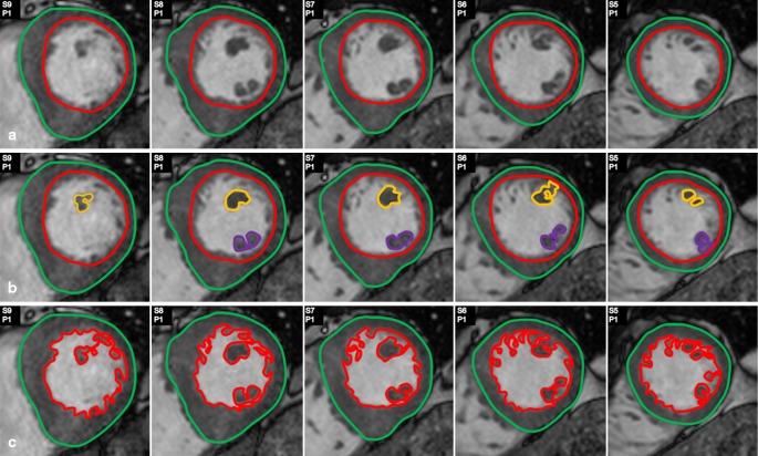
The use of cardiac magnetic resonance (CMR) analysis has increased in patients with hypertrophic cardiomyopathy (HCM). Quantification of left ventricular (LV) measures will be affected by the inclusion or exclusion of the papillary muscles as part of the LV mass, but the magnitude of effect and potential consequences are unknown.
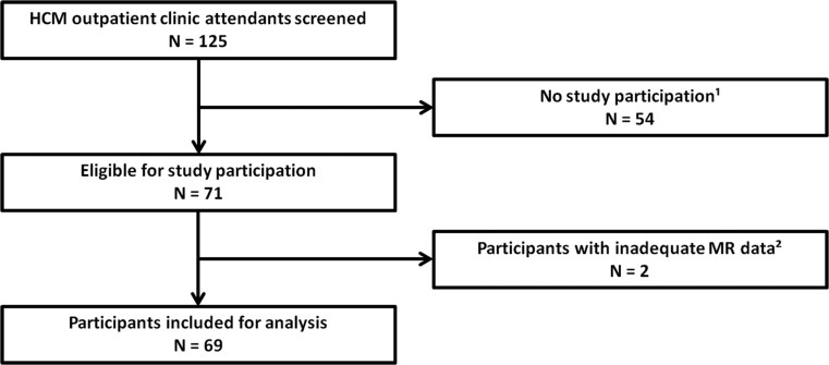
We performed Cine-CMR in (1) clinical HCM patients (n = 55) and (2) subclinical HCM mutation carriers without hypertrophy (n = 14). Absolute and relative differences in LV ejection fraction (EF) and mass were assessed between algorithms with and without inclusion of the papillary muscles.
Papillary muscle mass in group 1 was 6.6 ± 2.5 g/m(2) and inclusion of the papillary muscles resulted in significant relative increases in LVEF of 4.5 ± 1.8 % and in LV mass of 8.7 ± 2.6 %. For group 2 these figures were 4.0 ± 0.9 g/m(2), 3.8 ± 1.0 % and 9.5 ± 1.8 %, respectively. With a coefficient of variation of 4 %, this 9 % difference in LV mass during CMR follow-up will be considered a change, while in fact the exact same mass may have been assessed according to two different algorithms.
In clinical HCM patients, CMR quantification of important LV measures is significantly affected by inclusion or exclusion of the papillary muscles. In relative terms, the difference was similar in subjects without hypertrophy. This underscores a general need for a uniform approach in CMR image analysis.
肥厚型心肌病(HCM)患者中使用心脏磁共振(CMR)分析的情况有所增加。左心室(LV)测量的量化会受到将乳头肌纳入或排除在LV质量计算范围内的影响,但这种影响的程度和潜在后果尚不清楚。
我们对(1)临床HCM患者(n = 55)和(2)无肥厚的亚临床HCM突变携带者(n = 14)进行了电影CMR检查。评估了包含和不包含乳头肌的算法之间LV射血分数(EF)和质量的绝对和相对差异。
第1组的乳头肌质量为6.6±2.5 g/m²,将乳头肌纳入计算导致LVEF相对显著增加4.5±1.8%,LV质量相对显著增加8.7±2.6%。对于第2组,这些数字分别为4.0±0.9 g/m²、3.8±1.0%和9.5±1.8%。由于变异系数为4%,CMR随访期间LV质量9%的差异将被视为一种变化,而实际上根据两种不同算法评估的可能是完全相同的质量。
在临床HCM患者中,CMR对重要LV测量的量化受到乳头肌纳入或排除的显著影响。相对而言,在无肥厚的受试者中差异相似。这突出了CMR图像分析中统一方法的普遍需求。