• 文献检索
  • 文档翻译
  • 深度研究
  • 学术资讯
  • Suppr Zotero 插件Zotero 插件
  • 邀请有礼
  • 套餐&价格
  • 历史记录
应用&插件
Suppr Zotero 插件Zotero 插件浏览器插件Mac 客户端Windows 客户端微信小程序
定价
高级版会员购买积分包购买API积分包
服务
文献检索文档翻译深度研究API 文档MCP 服务
关于我们
关于 Suppr公司介绍联系我们用户协议隐私条款
关注我们

Suppr 超能文献

核心技术专利:CN118964589B侵权必究
粤ICP备2023148730 号-1Suppr @ 2026

文献检索

告别复杂PubMed语法,用中文像聊天一样搜索,搜遍4000万医学文献。AI智能推荐,让科研检索更轻松。

立即免费搜索

文件翻译

保留排版,准确专业,支持PDF/Word/PPT等文件格式,支持 12+语言互译。

免费翻译文档

深度研究

AI帮你快速写综述,25分钟生成高质量综述,智能提取关键信息,辅助科研写作。

立即免费体验

心脏磁共振成像中造影剂灌注的模拟:迈向无创心脏灌注定量的一步。

Simulation of the Perfusion of Contrast Agent Used in Cardiac Magnetic Resonance: A Step Toward Non-invasive Cardiac Perfusion Quantification.

作者信息

Alves João R, de Queiroz Rafael A B, Bär Markus, Dos Santos Rodrigo W

机构信息

Graduate Program in Computational Modeling, Universidade Federal de Juiz de Fora, Juiz de Fora, Brazil.

Department of Mathematical Modeling and Data Analysis, Physikalisch-Technische Bundesanstalt, Berlin, Germany.

出版信息

Front Physiol. 2019 Mar 14;10:177. doi: 10.3389/fphys.2019.00177. eCollection 2019.

DOI:10.3389/fphys.2019.00177
PMID:30949059
原文链接:https://pmc.ncbi.nlm.nih.gov/articles/PMC6436070/
Abstract

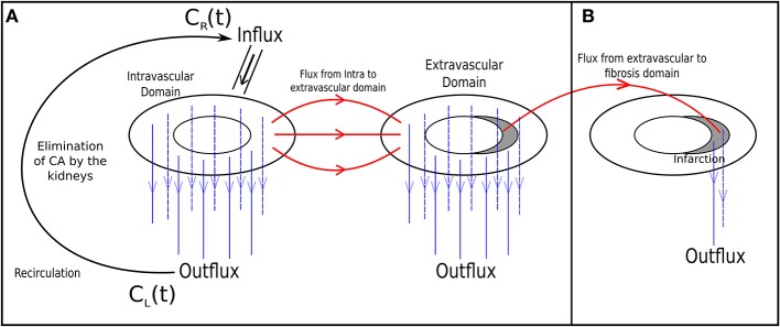
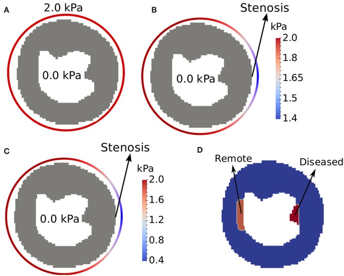
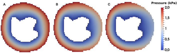
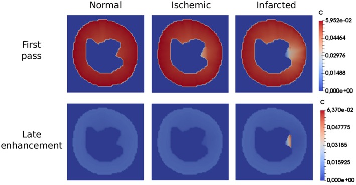
This work presents a new mathematical model to describe cardiac perfusion in the myocardium as acquired by cardiac magnetic resonance (CMR) perfusion exams. The combination of first pass (or contrast-enhanced CMR) and late enhancement CMR is a widely used non-invasive exam that can identify abnormal perfused regions of the heart via the use of a contrast agent (CA). The exam provides important information to the diagnosis, management, and prognosis of ischemia and infarct: perfusion on different regions, the status of microvascular structures, the presence of fibrosis, and the relative volume of extracellular space. This information is obtained by inferring the spatiotemporal dynamics of the contrast in the myocardial tissue from the acquired images. The evaluation of these physiological parameters plays an important role in the assessment of myocardial viability. However, the nature of cardiac physiology poses great challenges in the estimation of these parameters. Briefly, these are currently estimated qualitatively via visual inspection of images and comparison of relative brightness between different regions of the heart. Therefore, there is a great urge for techniques that can help to quantify cardiac perfusion. In this work, we propose a new mathematical model based on multidomain flow in porous media. The model is based on a system of partial differential equations. Darcy's law is used to obtain the pressure and velocity distribution. CA dynamics is described by reaction-diffusion-advection equations in the intravascular space and in the interstitial space. The interaction of fibrosis and the CA is also considered. The new model treats the domains as anisotropic media and imposes a closed loop of intravascular flow, which is necessary to reproduce the recirculation of the CA. The model parameters were adjusted to reproduce clinical data. In addition, the model was used to simulate different scenarios: normal perfusion; endocardial ischemia due to stenosis in a coronary artery in the epicardium; and myocardial infarct. Therefore, the computational model was able to correlate anatomical features, stenosis and the presence of fibrosis, with functional ones, cardiac perfusion. Altogether, the results suggest that the model can support the process of non-invasive cardiac perfusion quantification.

摘要

这项工作提出了一种新的数学模型,用于描述通过心脏磁共振(CMR)灌注检查获取的心肌中的心脏灌注情况。首次通过(或对比增强CMR)和延迟增强CMR的联合应用是一种广泛使用的非侵入性检查,可通过使用造影剂(CA)识别心脏的异常灌注区域。该检查为缺血和梗死的诊断、管理及预后提供重要信息:不同区域的灌注情况、微血管结构状态、纤维化的存在以及细胞外间隙的相对体积。这些信息是通过从获取的图像中推断心肌组织中造影剂的时空动态来获得的。这些生理参数的评估在心肌活力评估中起着重要作用。然而,心脏生理学的性质在这些参数的估计中带来了巨大挑战。简而言之,目前这些参数是通过对图像的目视检查以及心脏不同区域之间相对亮度的比较进行定性估计的。因此,迫切需要能够帮助量化心脏灌注的技术。在这项工作中,我们提出了一种基于多孔介质中多域流动的新数学模型。该模型基于偏微分方程组。利用达西定律获得压力和速度分布。造影剂动力学通过血管内空间和组织间隙空间中的反应 - 扩散 - 对流方程来描述。还考虑了纤维化与造影剂的相互作用。新模型将这些区域视为各向异性介质,并施加血管内流动的闭环,这对于再现造影剂的再循环是必要的。对模型参数进行了调整以再现临床数据。此外,该模型用于模拟不同场景:正常灌注;由于心外膜冠状动脉狭窄导致的心内膜缺血;以及心肌梗死。因此,该计算模型能够将解剖特征、狭窄和纤维化的存在与功能特征——心脏灌注联系起来。总之,结果表明该模型可以支持非侵入性心脏灌注量化过程。

https://cdn.ncbi.nlm.nih.gov/pmc/blobs/d73c/6436070/bf3ee4864b95/fphys-10-00177-g0012.jpg
https://cdn.ncbi.nlm.nih.gov/pmc/blobs/d73c/6436070/695a598d3fc8/fphys-10-00177-g0001.jpg
https://cdn.ncbi.nlm.nih.gov/pmc/blobs/d73c/6436070/aad2c8cc8e76/fphys-10-00177-g0002.jpg
https://cdn.ncbi.nlm.nih.gov/pmc/blobs/d73c/6436070/47991783d2c2/fphys-10-00177-g0003.jpg
https://cdn.ncbi.nlm.nih.gov/pmc/blobs/d73c/6436070/b8be3b31f177/fphys-10-00177-g0004.jpg
https://cdn.ncbi.nlm.nih.gov/pmc/blobs/d73c/6436070/f2efd76a57cd/fphys-10-00177-g0005.jpg
https://cdn.ncbi.nlm.nih.gov/pmc/blobs/d73c/6436070/b972141526ec/fphys-10-00177-g0006.jpg
https://cdn.ncbi.nlm.nih.gov/pmc/blobs/d73c/6436070/845e024a1185/fphys-10-00177-g0007.jpg
https://cdn.ncbi.nlm.nih.gov/pmc/blobs/d73c/6436070/1d32d4764240/fphys-10-00177-g0008.jpg
https://cdn.ncbi.nlm.nih.gov/pmc/blobs/d73c/6436070/345f47d6575a/fphys-10-00177-g0009.jpg
https://cdn.ncbi.nlm.nih.gov/pmc/blobs/d73c/6436070/beee45364466/fphys-10-00177-g0010.jpg
https://cdn.ncbi.nlm.nih.gov/pmc/blobs/d73c/6436070/bc9dd88da6a6/fphys-10-00177-g0011.jpg
https://cdn.ncbi.nlm.nih.gov/pmc/blobs/d73c/6436070/bf3ee4864b95/fphys-10-00177-g0012.jpg

相似文献

1
Simulation of the Perfusion of Contrast Agent Used in Cardiac Magnetic Resonance: A Step Toward Non-invasive Cardiac Perfusion Quantification.心脏磁共振成像中造影剂灌注的模拟:迈向无创心脏灌注定量的一步。
Front Physiol. 2019 Mar 14;10:177. doi: 10.3389/fphys.2019.00177. eCollection 2019.
2
[Myocardial microcirculation in humans--new approaches using MRI].[人类心肌微循环——磁共振成像的新方法]
Herz. 2003 Mar;28(2):74-81. doi: 10.1007/s00059-003-2451-6.
3
A Hybrid Model for Cardiac Perfusion: Coupling a Discrete Coronary Arterial Tree Model with a Continuous Porous-Media Flow Model of the Myocardium.一种用于心脏灌注的混合模型:将离散冠状动脉树模型与心肌连续多孔介质流动模型相结合。
Entropy (Basel). 2023 Aug 18;25(8):1229. doi: 10.3390/e25081229.
4
Italian Society of Cardiovascular Echography (SIEC) Consensus Conference on the state of the art of contrast echocardiography.意大利心血管超声学会(SIEC)关于超声造影技术现状的共识会议
Ital Heart J. 2004 Apr;5(4):309-34.
5
Characterization of microvascular dysfunction after acute myocardial infarction by cardiovascular magnetic resonance first-pass perfusion and late gadolinium enhancement imaging.通过心血管磁共振首过灌注和延迟钆增强成像对急性心肌梗死后微血管功能障碍的特征分析
J Cardiovasc Magn Reson. 2006;8(6):831-7. doi: 10.1080/10976640600778049.
6
Clinical applications of cardiac magnetic resonance imaging.心脏磁共振成像的临床应用
Minerva Cardioangiol. 2009 Jun;57(3):299-313.
7
Assessment of myocardial perfusion by magnetic resonance imaging.通过磁共振成像评估心肌灌注。
Herz. 1997 Feb;22(1):16-28. doi: 10.1007/BF03044567.
8
Myocardial first pass perfusion assessed by cardiac magnetic resonance and coronary microvascular dysfunction in women with angina and no obstructive coronary artery disease.心脏磁共振评估的心肌首过灌注和女性心绞痛但无阻塞性冠状动脉疾病的冠状动脉微血管功能障碍。
Scand J Clin Lab Invest. 2019 Jul;79(4):238-246. doi: 10.1080/00365513.2019.1587670. Epub 2019 Mar 20.
9
Magnetic resonance perfusion imaging in ischemic heart disease.缺血性心脏病的磁共振灌注成像
J Magn Reson Imaging. 1999 Sep;10(3):423-33. doi: 10.1002/(sici)1522-2586(199909)10:3<423::aid-jmri26>3.0.co;2-n.
10
A non-contrast CMR index for assessing myocardial fibrosis.一种用于评估心肌纤维化的非对比心脏磁共振成像指标。
Magn Reson Imaging. 2017 Oct;42:69-73. doi: 10.1016/j.mri.2017.04.012. Epub 2017 Apr 28.

引用本文的文献

1
A Hybrid Model for Cardiac Perfusion: Coupling a Discrete Coronary Arterial Tree Model with a Continuous Porous-Media Flow Model of the Myocardium.一种用于心脏灌注的混合模型:将离散冠状动脉树模型与心肌连续多孔介质流动模型相结合。
Entropy (Basel). 2023 Aug 18;25(8):1229. doi: 10.3390/e25081229.
2
A Poroelastic Approach for Modelling Myocardial Oedema in Acute Myocarditis.一种用于模拟急性心肌炎中心肌水肿的多孔弹性方法。
Front Physiol. 2022 Jul 4;13:888515. doi: 10.3389/fphys.2022.888515. eCollection 2022.
3
Prediction of myocardial blood flow under stress conditions by means of a computational model.

本文引用的文献

1
Fully automated, inline quantification of myocardial blood flow with cardiovascular magnetic resonance: repeatability of measurements in healthy subjects.使用心血管磁共振进行全自动、在线心肌血流定量:健康受试者测量的可重复性。
J Cardiovasc Magn Reson. 2018 Jul 9;20(1):48. doi: 10.1186/s12968-018-0462-y.
2
Fully quantitative cardiovascular magnetic resonance myocardial perfusion ready for clinical use: a comparison between cardiovascular magnetic resonance imaging and positron emission tomography.全面定量心血管磁共振心肌灌注成像准备临床应用:心血管磁共振成像与正电子发射断层扫描比较。
J Cardiovasc Magn Reson. 2017 Oct 19;19(1):78. doi: 10.1186/s12968-017-0388-9.
3
通过计算模型预测应激状态下的心肌血流。
Eur J Nucl Med Mol Imaging. 2022 May;49(6):1894-1905. doi: 10.1007/s00259-021-05667-8. Epub 2022 Jan 5.
A vessel length-based method to compute coronary fractional flow reserve from optical coherence tomography images.
一种基于血管长度从光学相干断层扫描图像计算冠状动脉血流储备分数的方法。
Biomed Eng Online. 2017 Jun 26;16(1):83. doi: 10.1186/s12938-017-0365-4.
4
Cardiac T1 mapping in congenital heart disease: bolus vs. infusion protocols for measurements of myocardial extracellular volume fraction.先天性心脏病中的心脏T1映射:用于测量心肌细胞外容积分数的团注与输注方案
Int J Cardiovasc Imaging. 2017 Dec;33(12):1961-1968. doi: 10.1007/s10554-017-1191-2. Epub 2017 Jun 15.
5
Myocardial perfusion cardiovascular magnetic resonance: optimized dual sequence and reconstruction for quantification.心肌灌注心血管磁共振成像:用于定量分析的优化双序列及重建方法
J Cardiovasc Magn Reson. 2017 Apr 7;19(1):43. doi: 10.1186/s12968-017-0355-5.
6
Impact of the papillary muscles on cardiac magnetic resonance image analysis of important left ventricular parameters in hypertrophic cardiomyopathy.乳头肌对肥厚型心肌病左心室重要参数心脏磁共振成像分析的影响。
Neth Heart J. 2016 May;24(5):326-31. doi: 10.1007/s12471-016-0805-y.
7
Cardiac magnetic resonance imaging for myocardial perfusion and diastolic function-reference control values for women.用于心肌灌注和舒张功能的心脏磁共振成像——女性的参考对照值
Cardiovasc Diagn Ther. 2016 Feb;6(1):78-86. doi: 10.3978/j.issn.2223-3652.2015.09.03.
8
Quality-of-Life and Economic Outcomes of Assessing Fractional Flow Reserve With Computed Tomography Angiography: PLATFORM.基于 CT 血管造影的血流储备分数评估的生活质量和经济结果:PLATFORM 研究。
J Am Coll Cardiol. 2015 Dec 1;66(21):2315-2323. doi: 10.1016/j.jacc.2015.09.051. Epub 2015 Oct 14.
9
Collateral circulation formation determines the characteristic profiles of contrast-enhanced MRI in the infarcted myocardium of pigs.侧支循环形成决定了猪梗死心肌对比增强磁共振成像的特征性表现。
Acta Pharmacol Sin. 2015 Apr;36(4):463-72. doi: 10.1038/aps.2014.158. Epub 2015 Mar 23.
10
A quantitative high resolution voxel-wise assessment of myocardial blood flow from contrast-enhanced first-pass magnetic resonance perfusion imaging: microsphere validation in a magnetic resonance compatible free beating explanted pig heart model.基于对比增强首次通过磁共振灌注成像的心肌血流定量高分辨率体素评估:在磁共振兼容的自由跳动离体猪心脏模型中的微球验证
Eur Heart J Cardiovasc Imaging. 2015 Oct;16(10):1082-92. doi: 10.1093/ehjci/jev023. Epub 2015 Mar 25.