Peleg Smadar, Dar Gali, Steinberg Nili, Masharawi Youssef, Hershkovitz Israel
Department of Anatomy and Anthropology, Sackler Faculty of Medicine, Tel Aviv University, 69978 Ramat Aviv, Tel Aviv, Israel ; Department of Physical Therapy, Zefat Academic College, Jerusalem Street 11, P.O. Box 160, 13206 Zefat, Israel.
Department of Anatomy and Anthropology, Sackler Faculty of Medicine, Tel Aviv University, 69978 Ramat Aviv, Tel Aviv, Israel ; Department of Physical Therapy, Faculty of Social Welfare and Health Studies, University of Haifa, Mount Carmel, 31905 Haifa, Israel.
Springerplus. 2016 Feb 20;5:141. doi: 10.1186/s40064-016-1772-x. eCollection 2016.
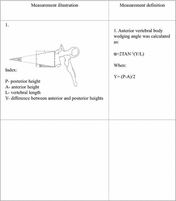
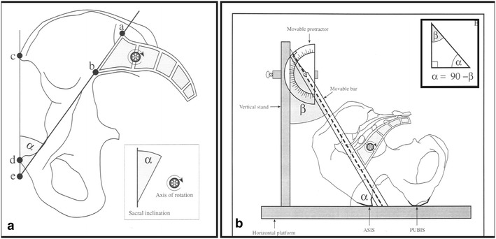
To examine whether the association between spinal alignment and sacral anatomical orientation (SAO) can be detected in skeletal populations, by comparing SAO values in individuals with a typical SD to individuals with normal spinal alignment. 2025 skeletons were screened for Scheuermann's disease. Scheuermann's kyphosis was established by the presence of apophyseal abnormalities associated with more than 5° of anterior wedging in each of three adjacent vertebrae. SAO was measured as the angle created between the intersection of a line running parallel to the superior surface of the sacrum and a line running between the anterior superior iliac spine and the anterior-superior edge of the symphysis pubis (PUBIS). SAO was measured on 185 individuals with normal spines and 183 individuals with Scheuermann's kyphosis. Out of 2025 skeletons, 183 (9 %) were diagnosed with Scheuermann's kyphosis. The sacrum was significantly more horizontally oriented in individuals with Scheuermann's kyphosis compared with the control (SAO: 44.44 ± 9.7° vs. 50 ± 9.9°, p < 0.001). Alteration in spinal biomechanics due to a horizontally orientated sacrum may be an important contributing factor for the development of Scheuermann's kyphosis.
为了通过比较典型脊柱滑脱个体与脊柱排列正常个体的骶骨解剖方位(SAO)值,来检验在骨骼人群中是否能检测到脊柱排列与骶骨解剖方位之间的关联。对2025具骨骼进行了休门氏病筛查。休门氏后凸畸形通过三个相邻椎体中每个椎体出现与超过5°的前楔形相关的骨骺异常来确定。SAO的测量方法是,一条平行于骶骨上表面的线与一条连接髂前上棘和耻骨联合前上缘的线的交点所形成的角度。对185名脊柱正常的个体和183名患有休门氏后凸畸形的个体进行了SAO测量。在2025具骨骼中,183具(9%)被诊断为休门氏后凸畸形。与对照组相比,患有休门氏后凸畸形的个体的骶骨明显更水平(SAO:44.44±9.7°对50±9.9°,p<0.001)。由于骶骨水平方向导致的脊柱生物力学改变可能是休门氏后凸畸形发展的一个重要促成因素。