• 文献检索
  • 文档翻译
  • 深度研究
  • 学术资讯
  • Suppr Zotero 插件Zotero 插件
  • 邀请有礼
  • 套餐&价格
  • 历史记录
应用&插件
Suppr Zotero 插件Zotero 插件浏览器插件Mac 客户端Windows 客户端微信小程序
定价
高级版会员购买积分包购买API积分包
服务
文献检索文档翻译深度研究API 文档MCP 服务
关于我们
关于 Suppr公司介绍联系我们用户协议隐私条款
关注我们

Suppr 超能文献

核心技术专利:CN118964589B侵权必究
粤ICP备2023148730 号-1Suppr @ 2026

文献检索

告别复杂PubMed语法,用中文像聊天一样搜索,搜遍4000万医学文献。AI智能推荐,让科研检索更轻松。

立即免费搜索

文件翻译

保留排版,准确专业,支持PDF/Word/PPT等文件格式,支持 12+语言互译。

免费翻译文档

深度研究

AI帮你快速写综述,25分钟生成高质量综述,智能提取关键信息,辅助科研写作。

立即免费体验

扩散加权成像和扩散张量成像在移植肾评估中的应用

Diffusion weighted imaging and diffusion tensor imaging in the evaluation of transplanted kidneys.

作者信息

Palmucci Stefano, Cappello Giuseppina, Attinà Giancarlo, Foti Pietro Valerio, Siverino Rita Olivia Anna, Roccasalva Federica, Piccoli Marina, Sinagra Nunziata, Milone Pietro, Veroux Massimiliano, Ettorre Giovanni Carlo

机构信息

Radiodiagnostic and Radiotherapy Unit, University Hospital "Policlinico-Vittorio Emanuele", Via Santa Sofia 78, 95123 Catania, Italy.

Vascular Surgery and Organ Transplant Unit, Department of Surgery, Transplantation and Advanced Technologies, University Hospital "Policlinico-Vittorio Emanuele", Via Santa Sofia 78, 95123 Catania, Italy.

出版信息

Eur J Radiol Open. 2015 May 16;2:71-80. doi: 10.1016/j.ejro.2015.05.001. eCollection 2015.

DOI:10.1016/j.ejro.2015.05.001
PMID:26937439
原文链接:https://pmc.ncbi.nlm.nih.gov/articles/PMC4750625/
Abstract

OBJECTIVE

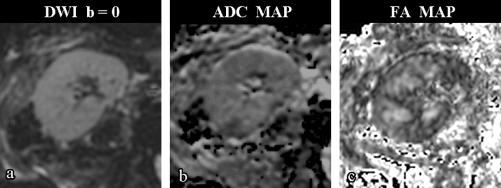
The aim of this study is to investigate the relation between renal indexes and functional MRI in a population of kidney transplant recipients who underwent MR with diffusion-weighted imaging (DWI) and diffusion tensor imaging (DTI) of the transplanted graft.

METHOD

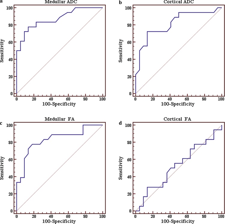
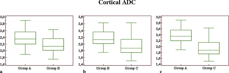
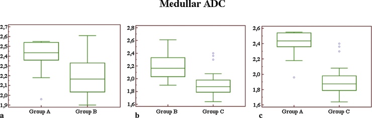
Study population included 40 patients with single kidney transplant. The patients were divided into 3 groups, on the basis of creatinine clearance (CrCl) values calculated using Cockcroft-Gault formula: group A, including patients with normal renal function (CrCl ≥ 60 mL/min); group B, which refers to patients with moderate renal impairment (CrCl > 30 but <60 mL/min); and, finally, group C, which means severe renal deterioration (CrCl ≤ 30 mL/min). All patients were investigated with a 1.5 Tesla MRI scanner, acquiring DWI and DTI sequences. A Mann-Whitney U test was adopted to compare apparent diffusion coefficients (ADCs) and fractional anisotropy (FA) measurements between groups. Receiver operating characteristic (ROC) curves were created for prediction of normal renal function (group A) and renal failure (group C). Pearson correlation was performed between renal clearance and functional imaging parameter (ADC and FA), obtained for cortical and medullar regions.

RESULTS

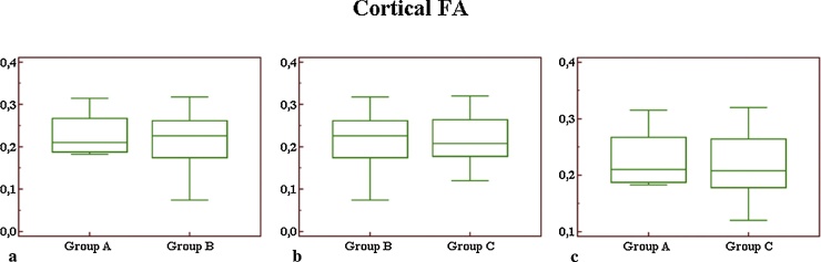
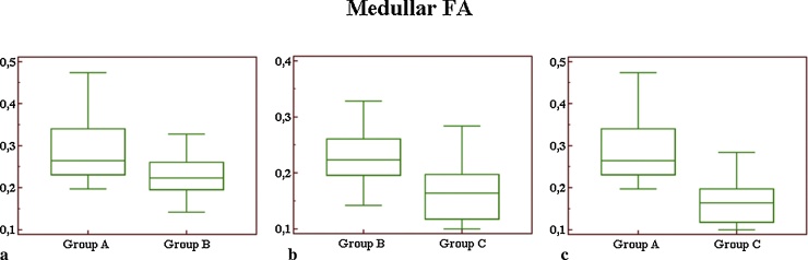
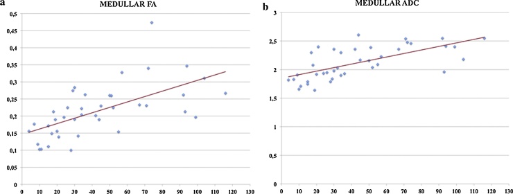
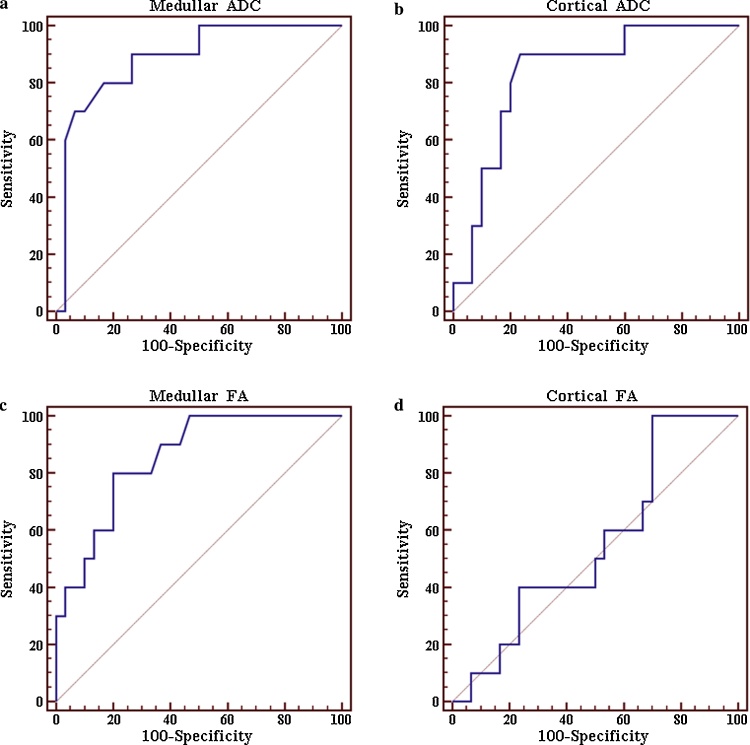
Mann-Whitney U test revealed a highly significant difference (p < 0.01) between patients with low CrCl (group C) and normal CrCl (group A) considering both medullar ADC and FA and cortical ADC. Regarding contiguous groups, the difference between group B and C was highly significant (p < 0.01) for medullar ADC and significant (p < 0.05) for cortical ADC and medullar FA. No difference between these groups was found considering cortical FA. Analyzing groups A and B, we found a significant difference (p < 0.05) for medullar both ADC and FA, while no difference was found for cortical ADC and FA. Strongest Pearson correlation was found between CrCl and medullar ADC (r = 0.65). For predicting normal renal function or severe renal impairment, highest values of AUC were observed using medullar ADC cut-off values (respectively 0.885 and 0.871); medullar FA showed also high accuracy (respectively 0.831 and 0.853).

CONCLUSIONS

DWI and DTI are promising tools for non-invasive monitoring of renal function; medullar ADC proved to be the best parameter for renal function assessment.

摘要

目的

本研究旨在调查接受移植肾磁共振成像(MRI)检查(包括扩散加权成像(DWI)和扩散张量成像(DTI))的肾移植受者群体中,肾脏指标与功能MRI之间的关系。

方法

研究人群包括40名单肾移植患者。根据使用Cockcroft - Gault公式计算的肌酐清除率(CrCl)值,将患者分为3组:A组,包括肾功能正常(CrCl≥60 mL/min)的患者;B组,指中度肾功能损害(CrCl>30但<60 mL/min)的患者;最后是C组,即严重肾功能恶化(CrCl≤30 mL/min)的患者。所有患者均使用1.5特斯拉MRI扫描仪进行检查,采集DWI和DTI序列。采用Mann - Whitney U检验比较各组之间的表观扩散系数(ADC)和分数各向异性(FA)测量值。绘制受试者操作特征(ROC)曲线以预测正常肾功能(A组)和肾衰竭(C组)。对皮质和髓质区域获得的肾脏清除率与功能成像参数(ADC和FA)进行Pearson相关性分析。

结果

Mann - Whitney U检验显示,考虑髓质ADC、FA以及皮质ADC时,低CrCl(C组)患者与正常CrCl(A组)患者之间存在高度显著差异(p<0.01)。对于相邻组,B组和C组之间在髓质ADC方面差异高度显著(p<0.01),在皮质ADC和髓质FA方面差异显著(p<0.05)。考虑皮质FA时,这些组之间未发现差异。分析A组和B组,我们发现髓质ADC和FA均存在显著差异(p<0.05),而皮质ADC和FA未发现差异。CrCl与髓质ADC之间发现最强的Pearson相关性(r = 0.65)。对于预测正常肾功能或严重肾功能损害,使用髓质ADC临界值时观察到最高的曲线下面积(AUC)值(分别为0.885和0.871);髓质FA也显示出较高的准确性(分别为0.831和0.853)。

结论

DWI和DTI是用于肾功能无创监测的有前景的工具;髓质ADC被证明是评估肾功能的最佳参数。

https://cdn.ncbi.nlm.nih.gov/pmc/blobs/a04b/4750625/64f856ef253e/gr8.jpg
https://cdn.ncbi.nlm.nih.gov/pmc/blobs/a04b/4750625/31c48aa11c5d/gr1.jpg
https://cdn.ncbi.nlm.nih.gov/pmc/blobs/a04b/4750625/cdebea4c7d0f/gr2.jpg
https://cdn.ncbi.nlm.nih.gov/pmc/blobs/a04b/4750625/3fffe7dc242d/gr3.jpg
https://cdn.ncbi.nlm.nih.gov/pmc/blobs/a04b/4750625/6e264e7b8185/gr4.jpg
https://cdn.ncbi.nlm.nih.gov/pmc/blobs/a04b/4750625/42602edebdbb/gr5.jpg
https://cdn.ncbi.nlm.nih.gov/pmc/blobs/a04b/4750625/5e350bde5d7c/gr6.jpg
https://cdn.ncbi.nlm.nih.gov/pmc/blobs/a04b/4750625/040ca8766473/gr7.jpg
https://cdn.ncbi.nlm.nih.gov/pmc/blobs/a04b/4750625/64f856ef253e/gr8.jpg

相似文献

1
Diffusion weighted imaging and diffusion tensor imaging in the evaluation of transplanted kidneys.扩散加权成像和扩散张量成像在移植肾评估中的应用
Eur J Radiol Open. 2015 May 16;2:71-80. doi: 10.1016/j.ejro.2015.05.001. eCollection 2015.
2
Diffusion-MR in kidney transplant recipients: is diuretic stimulation a useful diagnostic tool for improving differentiation between functioning and non-functioning kidneys?肾移植受者的扩散加权磁共振成像:利尿刺激是否是改善功能肾与无功能肾鉴别诊断的有用工具?
Clin Imaging. 2019 Jan-Feb;53:97-104. doi: 10.1016/j.clinimag.2018.10.003. Epub 2018 Oct 5.
3
Magnetic resonance with diffusion-weighted imaging in the evaluation of transplanted kidneys: preliminary findings.磁共振扩散加权成像在移植肾评估中的应用:初步研究结果。
Transplant Proc. 2011 May;43(4):960-6. doi: 10.1016/j.transproceed.2011.01.157.
4
Magnetic resonance with diffusion-weighted imaging in the evaluation of transplanted kidneys: updating results in 35 patients.磁共振扩散加权成像在移植肾评估中的应用:35例患者的最新结果
Transplant Proc. 2012 Sep;44(7):1884-8. doi: 10.1016/j.transproceed.2012.06.045.
5
Assessment of renal allograft function early after transplantation with isotropic resolution diffusion tensor imaging.移植后早期等分辨率扩散张量成像评估肾移植功能。
Eur Radiol. 2016 Feb;26(2):567-75. doi: 10.1007/s00330-015-3841-x. Epub 2015 May 28.
6
Comparison of the diagnostic performances of diffusion parameters in diffusion weighted imaging and diffusion tensor imaging of breast lesions.扩散加权成像和扩散张量成像中扩散参数对乳腺病变诊断性能的比较。
Eur J Radiol. 2013 Dec;82(12):e801-6. doi: 10.1016/j.ejrad.2013.09.001. Epub 2013 Sep 13.
7
Efficacy of diffusion tensor imaging in identification of degenerative cervical spondylotic myelopathy.弥散张量成像在诊断退行性颈椎脊髓病中的效能
Eur J Radiol Open. 2018 Dec 12;6:16-23. doi: 10.1016/j.ejro.2018.08.006. eCollection 2019.
8
Kidney transplant: functional assessment with diffusion-tensor MR imaging at 3T.肾脏移植:3T 磁共振扩散张量成像的功能评估。
Radiology. 2013 Jan;266(1):218-25. doi: 10.1148/radiol.12112522. Epub 2012 Nov 20.
9
Early assessment of acute kidney injury in severe acute pancreatitis with multimodal DWI: an animal model.多模态 DWI 对重症急性胰腺炎急性肾损伤的早期评估:动物模型研究。
Eur Radiol. 2023 Nov;33(11):7744-7755. doi: 10.1007/s00330-023-09782-y. Epub 2023 Jun 27.
10
Difference in Diffusion-Weighted Magnetic Resonance Imaging and Diffusion Tensor Imaging Parameters Between Endometrioid Endometrial Adenocarcinoma and Uterine Serous Adenocarcinoma: A Retrospective Study.子宫内膜样腺癌与子宫浆液性腺癌之间扩散加权磁共振成像和扩散张量成像参数的差异:一项回顾性研究
Int J Gynecol Cancer. 2017 Oct;27(8):1708-1713. doi: 10.1097/IGC.0000000000001054.

引用本文的文献

1
Diffusion Tensor Imaging (DTI) as a Non-Invasive Tool for Assessing Pediatric Kidney Transplants: A Feasibility Study.扩散张量成像(DTI)作为评估小儿肾移植的非侵入性工具:一项可行性研究。
Pediatr Transplant. 2025 Aug;29(5):e70119. doi: 10.1111/petr.70119.
2
Diffusion tensor imaging for the study of early renal dysfunction in patients affected by bardet-biedl syndrome.弥散张量成像在研究巴尔-杜-布综合征患者早期肾功能障碍中的应用。
Sci Rep. 2021 Oct 21;11(1):20855. doi: 10.1038/s41598-021-00394-4.
3
Hip Position Acutely Affects Oxygenation and Perfusion of Kidney Grafts as Measured by Functional Magnetic Resonance Imaging Methods-The Bent Knee Study.

本文引用的文献

1
Ultrasound-guided percutaneous renal biopsy in 295 children and adolescents: role of ultrasound and analysis of complications.295例儿童及青少年的超声引导下经皮肾活检:超声的作用及并发症分析
PLoS One. 2014 Dec 9;9(12):e114737. doi: 10.1371/journal.pone.0114737. eCollection 2014.
2
Chronic kidney disease: pathological and functional assessment with diffusion tensor imaging at 3T MR.慢性肾脏病:3T磁共振成像扩散张量成像的病理与功能评估
Eur Radiol. 2015 Mar;25(3):652-60. doi: 10.1007/s00330-014-3461-x. Epub 2014 Oct 11.
3
Assessment of renal fibrosis in chronic kidney disease using diffusion-weighted MRI.
通过功能磁共振成像方法测量发现,髋关节位置会急性影响肾移植的氧合和灌注——屈膝研究。
Front Med (Lausanne). 2021 Aug 10;8:697055. doi: 10.3389/fmed.2021.697055. eCollection 2021.
4
Renal Diffusion-Weighted Imaging (DWI) for Apparent Diffusion Coefficient (ADC), Intravoxel Incoherent Motion (IVIM), and Diffusion Tensor Imaging (DTI): Basic Concepts.肾脏弥散加权成像(DWI)的表观扩散系数(ADC)、体素内不相干运动(IVIM)和弥散张量成像(DTI):基本概念。
Methods Mol Biol. 2021;2216:187-204. doi: 10.1007/978-1-0716-0978-1_11.
5
Multiparametric Functional Magnetic Resonance Imaging for Evaluating Renal Allograft Injury.多参数功能磁共振成像评估肾移植损伤。
Korean J Radiol. 2019 Jun;20(6):894-908. doi: 10.3348/kjr.2018.0540.
6
Renal Damages in Deoxycorticosterone Acetate-Salt Hypertensive Rats: Assessment with Diffusion Tensor Imaging and T2-mapping.去氧皮质酮醋酸盐高血压大鼠的肾脏损伤:扩散张量成像和 T2 映射评估。
Mol Imaging Biol. 2020 Feb;22(1):94-104. doi: 10.1007/s11307-019-01364-z.
7
Change in kidney volume after kidney transplantation in patients with autosomal polycystic kidney disease.自体多囊肾病患者肾移植后肾脏体积的变化。
PLoS One. 2018 Dec 27;13(12):e0209332. doi: 10.1371/journal.pone.0209332. eCollection 2018.
8
Diffusion-weighted magnetic resonance imaging to assess diffuse renal pathology: a systematic review and statement paper.磁共振弥散加权成像评估弥漫性肾脏病变:系统评价和立场声明。
Nephrol Dial Transplant. 2018 Sep 1;33(suppl_2):ii29-ii40. doi: 10.1093/ndt/gfy163.
9
Scintigraphic texture analysis for assessment of renal allograft function.用于评估肾移植功能的闪烁扫描纹理分析
Pol J Radiol. 2018 Jan 18;83:e1-e10. doi: 10.5114/pjr.2018.74956. eCollection 2018.
10
Fast diffusion kurtosis imaging of fibrotic mouse kidneys.纤维化小鼠肾脏的快速扩散峰度成像
NMR Biomed. 2016 Dec;29(12):1709-1719. doi: 10.1002/nbm.3623. Epub 2016 Oct 12.
使用扩散加权磁共振成像评估慢性肾脏病中的肾纤维化
Clin Radiol. 2014 Nov;69(11):1117-22. doi: 10.1016/j.crad.2014.06.011. Epub 2014 Jul 23.
4
Renal function, nephrogenic systemic fibrosis and other adverse reactions associated with gadolinium-based contrast media.肾功能、肾源性系统性纤维化及与钆基造影剂相关的其他不良反应。
Nefrologia. 2014;34(4):428-38. doi: 10.3265/Nefrologia.pre2014.Apr.12375.
5
MR diffusion tensor imaging of normal kidneys.正常肾脏的磁共振扩散张量成像
J Magn Reson Imaging. 2014 Nov;40(5):1099-102. doi: 10.1002/jmri.24450. Epub 2013 Oct 29.
6
3T magnetic resonance diffusion tensor imaging in chronic kidney disease.3T磁共振扩散张量成像在慢性肾脏病中的应用
Abdom Imaging. 2014 Aug;39(4):770-5. doi: 10.1007/s00261-014-0116-y.
7
Diffusion Tensor Imaging of the Kidneys: Influence of b-Value and Number of Encoding Directions on Image Quality and Diffusion Tensor Parameters.肾脏的扩散张量成像:b值和编码方向数量对图像质量及扩散张量参数的影响
J Clin Imaging Sci. 2013 Nov 28;3:53. doi: 10.4103/2156-7514.122323. eCollection 2013.
8
Diffusion-weighted imaging and apparent diffusion coefficient values versus contrast-enhanced MR imaging in the identification and characterisation of acute pyelonephritis.扩散加权成像和表观扩散系数值与对比增强磁共振成像在急性肾盂肾炎的识别和特征化中的比较。
Eur Radiol. 2013 Dec;23(12):3501-8. doi: 10.1007/s00330-013-2951-6. Epub 2013 Jul 26.
9
Diffusion tensor parameters and principal eigenvector coherence: relation to b-value intervals and field strength.弥散张量参数和主特征向量相干性:与 b 值间隔和场强的关系。
Magn Reson Imaging. 2013 Jun;31(5):742-7. doi: 10.1016/j.mri.2012.11.014. Epub 2013 Jan 30.
10
Diffusion tensor imaging and tractography of the kidneys: assessment of chronic parenchymal diseases.肾脏弥散张量成像和纤维束追踪:慢性实质疾病评估。
Eur Radiol. 2013 Jun;23(6):1678-85. doi: 10.1007/s00330-012-2749-y. Epub 2013 Jan 9.