• 文献检索
  • 文档翻译
  • 深度研究
  • 学术资讯
  • Suppr Zotero 插件Zotero 插件
  • 邀请有礼
  • 套餐&价格
  • 历史记录
应用&插件
Suppr Zotero 插件Zotero 插件浏览器插件Mac 客户端Windows 客户端微信小程序
定价
高级版会员购买积分包购买API积分包
服务
文献检索文档翻译深度研究API 文档MCP 服务
关于我们
关于 Suppr公司介绍联系我们用户协议隐私条款
关注我们

Suppr 超能文献

核心技术专利:CN118964589B侵权必究
粤ICP备2023148730 号-1Suppr @ 2026

文献检索

告别复杂PubMed语法,用中文像聊天一样搜索,搜遍4000万医学文献。AI智能推荐,让科研检索更轻松。

立即免费搜索

文件翻译

保留排版,准确专业,支持PDF/Word/PPT等文件格式,支持 12+语言互译。

免费翻译文档

深度研究

AI帮你快速写综述,25分钟生成高质量综述,智能提取关键信息,辅助科研写作。

立即免费体验

用于检测卵巢组织中乳腺癌细胞的细胞表面标志物的鉴定

Identification of cell-surface markers for detecting breast cancer cells in ovarian tissue.

作者信息

Peters Inge T A, Hilders Carina G J M, Sier Cornelis F M, Vahrmeijer Alexander L, Smit Vincent T H B M, Baptist Trimbos J, Kuppen Peter J K

机构信息

Department of Gynecology, Leiden University Medical Center, Leiden, The Netherlands.

Department of Gynecology, Reinier de Graaf Hospital, Delft, The Netherlands.

出版信息

Arch Gynecol Obstet. 2016 Aug;294(2):385-93. doi: 10.1007/s00404-016-4036-7. Epub 2016 Mar 5.

DOI:10.1007/s00404-016-4036-7
PMID:26946151
原文链接:https://pmc.ncbi.nlm.nih.gov/articles/PMC4937067/
Abstract

PURPOSE

The safety of ovarian tissue autotransplantation in oncology patients cannot be ensured, as current tumor-detection methods compromise the ovarian tissue viability. Although non-destructive methods (for instance near-infrared fluorescence imaging) can discriminate malignant from healthy tissues while leaving the examined tissues unaffected, they require specific cell-surface tumor markers. We determined which tumor markers are suitable targets for tumor-specific imaging to exclude the presence of breast cancer cells in ovarian tissue.

METHODS

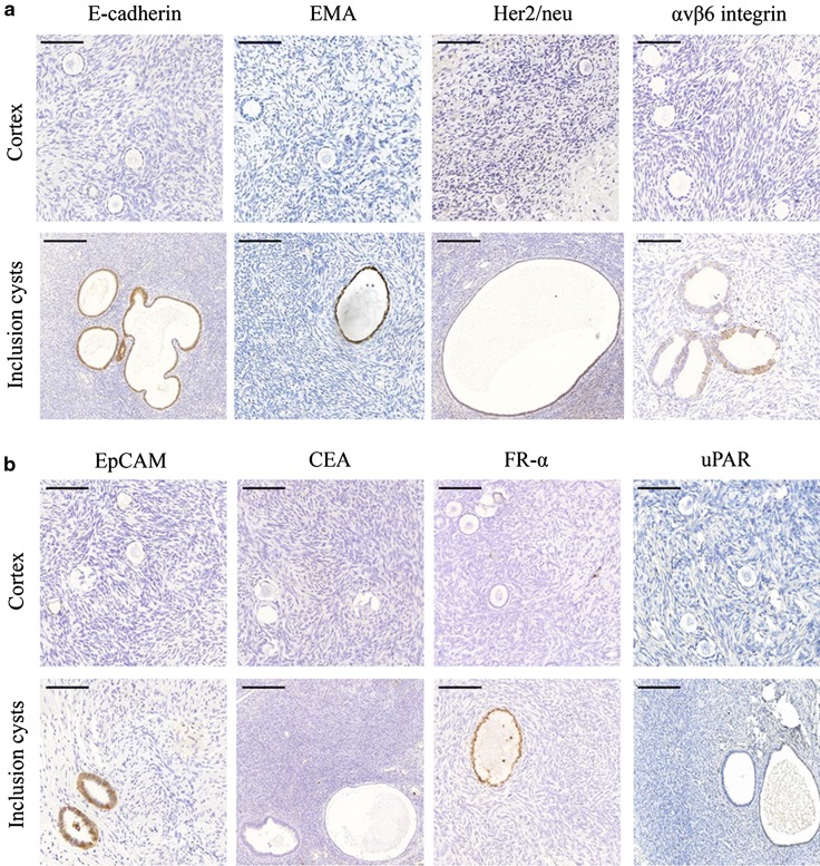
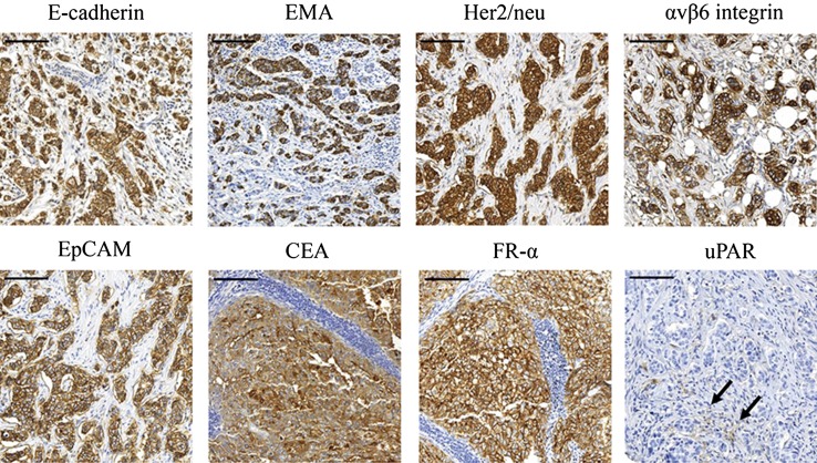
Immunohistochemistry was performed on formalin-fixed, paraffin-embedded specimens of ten ovaries from premenopausal patients. Additionally, we screened a tissue microarray containing tumor tissue cores from 24 breast cancer patients being eligible for ovarian tissue cryopreservation. The following cell-surface tumor markers were tested: E-cadherin, EMA (epithelial membrane antigen), Her2/neu (human epidermal growth factor receptor type 2), αvβ6 integrin, EpCAM (epithelial cell adhesion molecule), CEA (carcinoembryonic antigen), FR-α (folate receptor-alpha), and uPAR (urokinase-type plasminogen activator receptor). For each tumor, the percentage of positive breast tumor cells was measured.

RESULTS

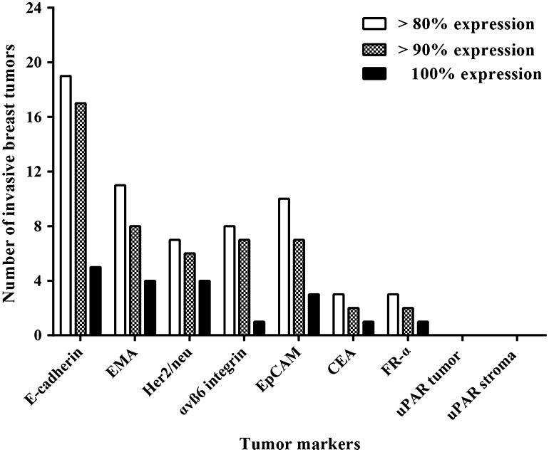
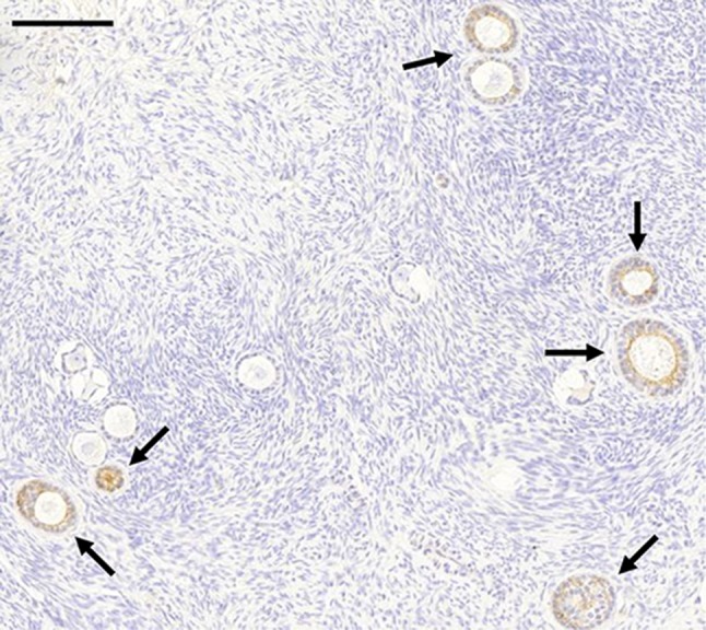
None of the ten ovaries were positive for any of the markers tested. However, all markers (except CEA and uPAR) were present on epithelial cells of inclusion cysts. E-cadherin was present in the majority of breast tumors: ≥90 % of tumor cells were positive for E-cadherin in 17 out of 24 tumors, and 100 % of tumor cells were positive in 5 out of 24 tumors.

CONCLUSIONS

Of the markers tested, E-cadherin is the most suitable marker for a tumor-specific probe in ovarian tissue. Methods are required to distinguish inclusion cysts from breast tumor cells.

摘要

目的

肿瘤患者卵巢组织自体移植的安全性无法得到保证,因为目前的肿瘤检测方法会损害卵巢组织的活力。尽管非破坏性方法(如近红外荧光成像)能够在不影响被检查组织的情况下区分恶性组织和健康组织,但它们需要特定的细胞表面肿瘤标志物。我们确定了哪些肿瘤标志物是肿瘤特异性成像的合适靶点,以排除卵巢组织中乳腺癌细胞的存在。

方法

对来自绝经前患者的10个卵巢的福尔马林固定、石蜡包埋标本进行免疫组织化学检测。此外,我们筛选了一个组织芯片,其中包含24例符合卵巢组织冷冻保存条件的乳腺癌患者的肿瘤组织芯。检测了以下细胞表面肿瘤标志物:E-钙黏蛋白、上皮膜抗原(EMA)、人表皮生长因子受体2(Her2/neu)、αvβ6整合素、上皮细胞黏附分子(EpCAM)、癌胚抗原(CEA)、叶酸受体α(FR-α)和尿激酶型纤溶酶原激活剂受体(uPAR)。对于每个肿瘤,测量阳性乳腺肿瘤细胞的百分比。

结果

所检测的10个卵巢中,没有一个对任何检测的标志物呈阳性。然而,所有标志物(CEA和uPAR除外)均存在于包涵囊肿的上皮细胞中。E-钙黏蛋白存在于大多数乳腺肿瘤中:24个肿瘤中有17个肿瘤中≥90%的肿瘤细胞E-钙黏蛋白呈阳性,24个肿瘤中有5个肿瘤中100%的肿瘤细胞呈阳性。

结论

在所检测的标志物中,E-钙黏蛋白是卵巢组织中肿瘤特异性探针最适合的标志物。需要有方法来区分包涵囊肿和乳腺肿瘤细胞。

https://cdn.ncbi.nlm.nih.gov/pmc/blobs/ca53/4937067/3fc07bebbdf5/404_2016_4036_Fig4_HTML.jpg
https://cdn.ncbi.nlm.nih.gov/pmc/blobs/ca53/4937067/795384515fc6/404_2016_4036_Fig1_HTML.jpg
https://cdn.ncbi.nlm.nih.gov/pmc/blobs/ca53/4937067/eb0c983effc2/404_2016_4036_Fig2_HTML.jpg
https://cdn.ncbi.nlm.nih.gov/pmc/blobs/ca53/4937067/a2fddea7fe28/404_2016_4036_Fig3_HTML.jpg
https://cdn.ncbi.nlm.nih.gov/pmc/blobs/ca53/4937067/3fc07bebbdf5/404_2016_4036_Fig4_HTML.jpg

相似文献

1
Identification of cell-surface markers for detecting breast cancer cells in ovarian tissue.用于检测卵巢组织中乳腺癌细胞的细胞表面标志物的鉴定
Arch Gynecol Obstet. 2016 Aug;294(2):385-93. doi: 10.1007/s00404-016-4036-7. Epub 2016 Mar 5.
2
Morphological and phenotypical features of ovarian metastases in breast cancer patients.乳腺癌患者卵巢转移灶的形态学和表型特征。
BMC Cancer. 2017 Mar 21;17(1):206. doi: 10.1186/s12885-017-3191-y.
3
New methods to improve the safety assessment of cryopreserved ovarian tissue for fertility preservation in breast cancer patients.改善乳腺癌患者生育力保存中冷冻卵巢组织安全性评估的新方法。
Fertil Steril. 2015 Dec;104(6):1493-502.e1-2. doi: 10.1016/j.fertnstert.2015.08.009. Epub 2015 Oct 1.
4
Profiling signalling pathways in formalin-fixed and paraffin-embedded breast cancer tissues reveals cross-talk between EGFR, HER2, HER3 and uPAR.在福尔马林固定和石蜡包埋的乳腺癌组织中分析信号通路,揭示了 EGFR、HER2、HER3 和 uPAR 之间的串扰。
J Cell Physiol. 2012 Jan;227(1):204-12. doi: 10.1002/jcp.22718.
5
Tumor stroma is the predominant uPA-, uPAR-, PAI-1-expressing tissue in human breast cancer: prognostic impact.肿瘤基质是人类乳腺癌中主要表达尿激酶型纤溶酶原激活物(uPA)、尿激酶型纤溶酶原激活物受体(uPAR)和纤溶酶原激活物抑制剂-1(PAI-1)的组织:预后影响。
Histol Histopathol. 2009 Jul;24(7):869-77. doi: 10.14670/HH-24.869.
6
Evaluation of quantitative polymerase chain reaction markers for the detection of breast cancer cells in ovarian tissue stored for fertility preservation.评估用于检测储存于生育力保存卵巢组织中乳腺癌细胞的定量聚合酶链反应标志物。
Fertil Steril. 2015 Aug;104(2):410-7.e4. doi: 10.1016/j.fertnstert.2015.04.036. Epub 2015 Jun 3.
7
Effect of macrophages on breast cancer cell proliferation, and on expression of hormone receptors, uPAR and HER-2.巨噬细胞对乳腺癌细胞增殖以及激素受体、uPAR 和 HER-2 表达的影响。
Int J Oncol. 2017 Jul;51(1):104-114. doi: 10.3892/ijo.2017.3996. Epub 2017 May 11.
8
Detection of urokinase plasminogen activator receptor and c-erbB-2 in sera of patients with breast and ovarian carcinoma.乳腺癌和卵巢癌患者血清中尿激酶型纤溶酶原激活物受体及c-erbB-2的检测
Clin Biochem. 2003 Oct;36(7):537-43. doi: 10.1016/s0009-9120(03)00102-4.
9
Correlation of E-cadherin expression and routine immunohistochemistry panel in breast invasive ductal carcinoma.乳腺浸润性导管癌中E-钙黏蛋白表达与常规免疫组化指标的相关性
Cancer Biomark. 2009;5(1):1-8. doi: 10.3233/CBM-2009-0551.
10
Expression of urokinase plasminogen activator and its receptor in advanced epithelial ovarian cancer patients.尿激酶型纤溶酶原激活剂及其受体在晚期上皮性卵巢癌患者中的表达
Gynecol Oncol. 2009 Aug;114(2):265-72. doi: 10.1016/j.ygyno.2009.04.031. Epub 2009 May 17.

引用本文的文献

1
Stepping forward: T-cell redirecting bispecific antibodies in cancer therapy.向前迈进:癌症治疗中重定向T细胞的双特异性抗体
Acta Pharm Sin B. 2024 Jun;14(6):2361-2377. doi: 10.1016/j.apsb.2024.03.027. Epub 2024 Mar 24.
2
Targeting Glycans and Heavily Glycosylated Proteins for Tumor Imaging.靶向聚糖和高度糖基化蛋白用于肿瘤成像
Cancers (Basel). 2020 Dec 21;12(12):3870. doi: 10.3390/cancers12123870.
3
Dual-Color Fluorescence Imaging of EpCAM and EGFR in Breast Cancer Cells with a Bull's Eye-Type Plasmonic Chip.利用牛眼型等离子体芯片对乳腺癌细胞中的 EpCAM 和 EGFR 进行双色荧光成像。

本文引用的文献

1
Searching for metastases in ovarian tissue before autotransplantation: a tailor-made approach.自体移植前在卵巢组织中寻找转移灶:一种量身定制的方法。
Fertil Steril. 2015 Feb;103(2):469-77. doi: 10.1016/j.fertnstert.2014.11.001. Epub 2014 Dec 10.
2
Fertility preservation for age-related fertility decline.与年龄相关的生育能力下降的生育力保存。
Lancet. 2014 Oct 4;384(9950):1311-9. doi: 10.1016/S0140-6736(14)61261-7.
3
Imaging of Mohs micrographic surgery sections using full-field optical coherence tomography: a pilot study.应用全场光学相干断层扫描技术对 Mohs 显微描记手术切片进行成像:一项初步研究。
Sensors (Basel). 2017 Dec 19;17(12):2942. doi: 10.3390/s17122942.
4
Morphological and phenotypical features of ovarian metastases in breast cancer patients.乳腺癌患者卵巢转移灶的形态学和表型特征。
BMC Cancer. 2017 Mar 21;17(1):206. doi: 10.1186/s12885-017-3191-y.
5
Prevalence and Risk Factors of Ovarian Metastases in Breast Cancer Patients < 41 Years of Age in the Netherlands: A Nationwide Retrospective Cohort Study.荷兰41岁以下乳腺癌患者卵巢转移的患病率及危险因素:一项全国性回顾性队列研究
PLoS One. 2017 Jan 26;12(1):e0168277. doi: 10.1371/journal.pone.0168277. eCollection 2017.
6
Selecting Targets for Tumor Imaging: An Overview of Cancer-Associated Membrane Proteins.肿瘤成像靶点的选择:癌症相关膜蛋白概述
Biomark Cancer. 2016 Sep 27;8:119-133. doi: 10.4137/BIC.S38542. eCollection 2016.
Dermatol Surg. 2014 Mar;40(3):266-74. doi: 10.1111/dsu.12419. Epub 2014 Jan 16.
4
Is transplantation of cryopreserved ovarian tissue from patients with advanced-stage breast cancer safe? A pilot study.晚期乳腺癌患者冷冻卵巢组织移植安全吗?一项初步研究。
J Assist Reprod Genet. 2013 Oct;30(10):1289-99. doi: 10.1007/s10815-013-0065-3. Epub 2013 Aug 29.
5
Image-guided cancer surgery using near-infrared fluorescence.近红外荧光引导的癌症手术。
Nat Rev Clin Oncol. 2013 Sep;10(9):507-18. doi: 10.1038/nrclinonc.2013.123. Epub 2013 Jul 23.
6
Autotransplantation of cryopreserved ovarian tissue in cancer survivors and the risk of reintroducing malignancy: a systematic review.癌症幸存者冷冻卵巢组织的自体移植与恶性肿瘤再引入风险:系统评价。
Hum Reprod Update. 2013 Sep-Oct;19(5):483-506. doi: 10.1093/humupd/dmt020. Epub 2013 Jun 30.
7
Near-infrared fluorescence-guided resection of colorectal liver metastases.近红外荧光引导下结直肠肝转移瘤切除术。
Cancer. 2013 Sep 15;119(18):3411-8. doi: 10.1002/cncr.28203. Epub 2013 Jun 21.
8
Restoration of ovarian activity and pregnancy after transplantation of cryopreserved ovarian tissue: a review of 60 cases of reimplantation.冷冻卵巢组织移植后卵巢功能和妊娠的恢复:60 例再植入的回顾性研究。
Fertil Steril. 2013 May;99(6):1503-13. doi: 10.1016/j.fertnstert.2013.03.030.
9
EpCAM: structure and function in health and disease.上皮细胞黏附分子(EpCAM):健康与疾病中的结构和功能
Biochim Biophys Acta. 2013 Aug;1828(8):1989-2001. doi: 10.1016/j.bbamem.2013.04.018. Epub 2013 Apr 23.
10
Risk of transferring malignant cells with transplanted frozen-thawed ovarian tissue.移植冷冻解冻卵巢组织转移恶性细胞的风险。
Fertil Steril. 2013 May;99(6):1514-22. doi: 10.1016/j.fertnstert.2013.03.027. Epub 2013 Mar 29.