Peters Inge T A, van der Steen Merle A, Huisman Bertine W, Hilders Carina G J M, Smit Vincent T H B M, Vahrmeijer Alexander L, Sier Cornelis F M, Trimbos J Baptist, Kuppen Peter J K
Department of Gynecology, Leiden University Medical Center, Leiden, the Netherlands.
Department of Surgery, Leiden University Medical Center, Leiden, the Netherlands.
BMC Cancer. 2017 Mar 21;17(1):206. doi: 10.1186/s12885-017-3191-y.
Autotransplantation of frozen-thawed ovarian tissue is a method to preserve ovarian function and fertility in patients undergoing gonadotoxic therapy. In oncology patients, the safety cannot yet be guaranteed, since current tumor detection methods can only exclude the presence of malignant cells in ovarian fragments that are not transplanted. We determined the need for a novel detection method by studying the distribution of tumor cells in ovaries from patients with breast cancer. Furthermore, we examined which cell-surface proteins are suitable as a target for non-invasive tumor-specific imaging of ovarian metastases from invasive breast cancer.
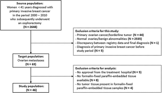
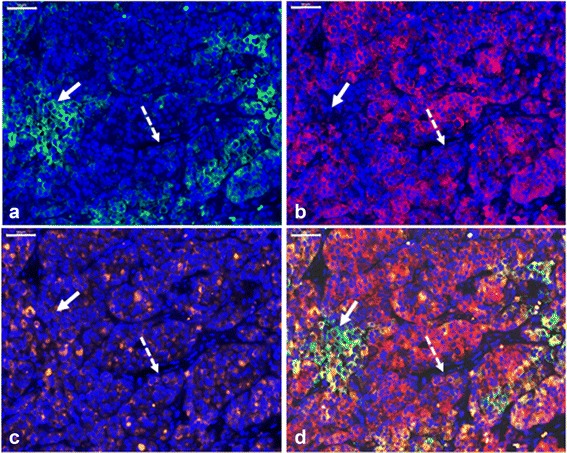
Using the nationwide database of the Dutch Pathology Registry (PALGA), we identified a cohort of 46 women with primary invasive breast cancer and ovarian metastases. The localization and morphology of ovarian metastases were determined on hematoxylin-and-eosin-stained sections. The following cell-surface markers were immunohistochemically analyzed: E-cadherin, epithelial membrane antigen (EMA), human epidermal growth receptor type 2 (Her2/neu), carcinoembryonic antigen (CEA), αvβ6 integrin and epithelial cell adhesion molecule (EpCAM).
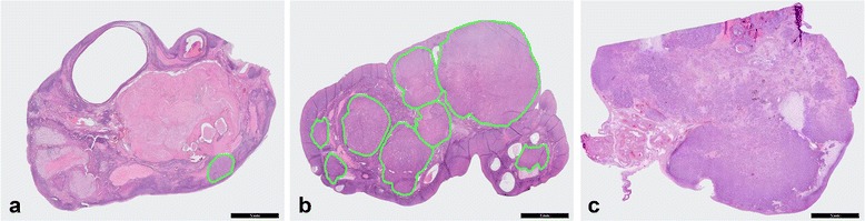
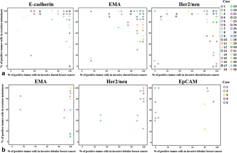
The majority of ovarian metastases (71%) consisted of a solitary metastasis or multiple distinct nodules separated by uninvolved ovarian tissue, suggesting that ovarian metastases might be overlooked by the current detection approach. Combining the targets E-cadherin, EMA and Her2/neu resulted in nearly 100% detection of ductal ovarian metastases, whereas the combination of EMA, Her2/neu and EpCAM was most suitable to detect lobular ovarian metastases.
Examination of the actual ovarian transplants is recommended. A combination of targets is most appropriate to detect ovarian metastases by tumor-specific imaging.
冻融卵巢组织自体移植是一种在接受性腺毒性治疗的患者中保留卵巢功能和生育能力的方法。在肿瘤患者中,安全性尚无法保证,因为目前的肿瘤检测方法只能排除未移植的卵巢组织碎片中恶性细胞的存在。我们通过研究乳腺癌患者卵巢中肿瘤细胞的分布,确定了对一种新型检测方法的需求。此外,我们还研究了哪些细胞表面蛋白适合作为浸润性乳腺癌卵巢转移灶非侵入性肿瘤特异性成像的靶点。
利用荷兰病理登记处(PALGA)的全国性数据库,我们确定了一组46例原发性浸润性乳腺癌伴卵巢转移的女性患者。在苏木精和伊红染色切片上确定卵巢转移灶的定位和形态。对以下细胞表面标志物进行免疫组织化学分析:E-钙黏蛋白、上皮膜抗原(EMA)、人表皮生长受体2型(Her2/neu)、癌胚抗原(CEA)、αvβ6整合素和上皮细胞黏附分子(EpCAM)。
大多数卵巢转移灶(71%)由单个转移灶或多个由未受累卵巢组织分隔的不同结节组成,这表明目前的检测方法可能会忽略卵巢转移灶。联合使用E-钙黏蛋白、EMA和Her2/neu靶点,几乎能100%检测到导管性卵巢转移灶,而EMA、Her2/neu和EpCAM的联合使用最适合检测小叶性卵巢转移灶。
建议对实际的卵巢移植组织进行检查。联合使用多个靶点最适合通过肿瘤特异性成像检测卵巢转移灶。