• 文献检索
  • 文档翻译
  • 深度研究
  • 学术资讯
  • Suppr Zotero 插件Zotero 插件
  • 邀请有礼
  • 套餐&价格
  • 历史记录
应用&插件
Suppr Zotero 插件Zotero 插件浏览器插件Mac 客户端Windows 客户端微信小程序
定价
高级版会员购买积分包购买API积分包
服务
文献检索文档翻译深度研究API 文档MCP 服务
关于我们
关于 Suppr公司介绍联系我们用户协议隐私条款
关注我们

Suppr 超能文献

核心技术专利:CN118964589B侵权必究
粤ICP备2023148730 号-1Suppr @ 2026

文献检索

告别复杂PubMed语法,用中文像聊天一样搜索,搜遍4000万医学文献。AI智能推荐,让科研检索更轻松。

立即免费搜索

文件翻译

保留排版,准确专业,支持PDF/Word/PPT等文件格式,支持 12+语言互译。

免费翻译文档

深度研究

AI帮你快速写综述,25分钟生成高质量综述,智能提取关键信息,辅助科研写作。

立即免费体验

用于评估中度年龄相关性黄斑变性中局灶性色素沉着变化的自动视网膜图像分析

Automated Retinal Image Analysis for Evaluation of Focal Hyperpigmentary Changes in Intermediate Age-Related Macular Degeneration.

作者信息

Schmitz-Valckenberg Steffen, Göbel Arno P, Saur Stefan C, Steinberg Julia S, Thiele Sarah, Wojek Christian, Russmann Christoph, Holz Frank G

机构信息

Department of Ophthalmology, University of Bonn, Bonn, Germany.

Carl Zeiss AG, Oberkochen, Germany.

出版信息

Transl Vis Sci Technol. 2016 Mar 4;5(2):3. doi: 10.1167/tvst.5.2.3. eCollection 2016 Mar.

DOI:10.1167/tvst.5.2.3
PMID:26966639
原文链接:https://pmc.ncbi.nlm.nih.gov/articles/PMC4782823/
Abstract

PURPOSE

To develop and evaluate a software tool for automated detection of focal hyperpigmentary changes (FHC) in eyes with intermediate age-related macular degeneration (AMD).

METHODS

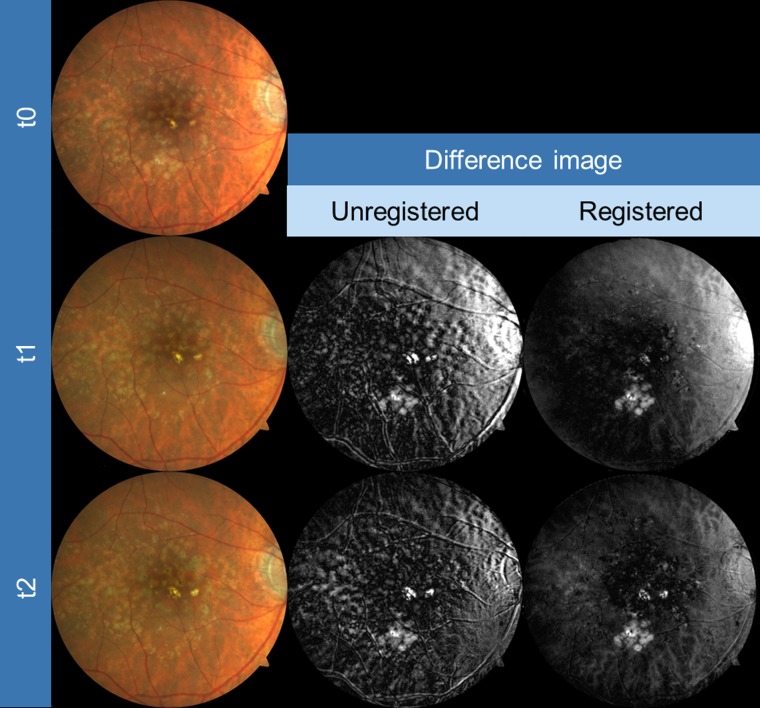
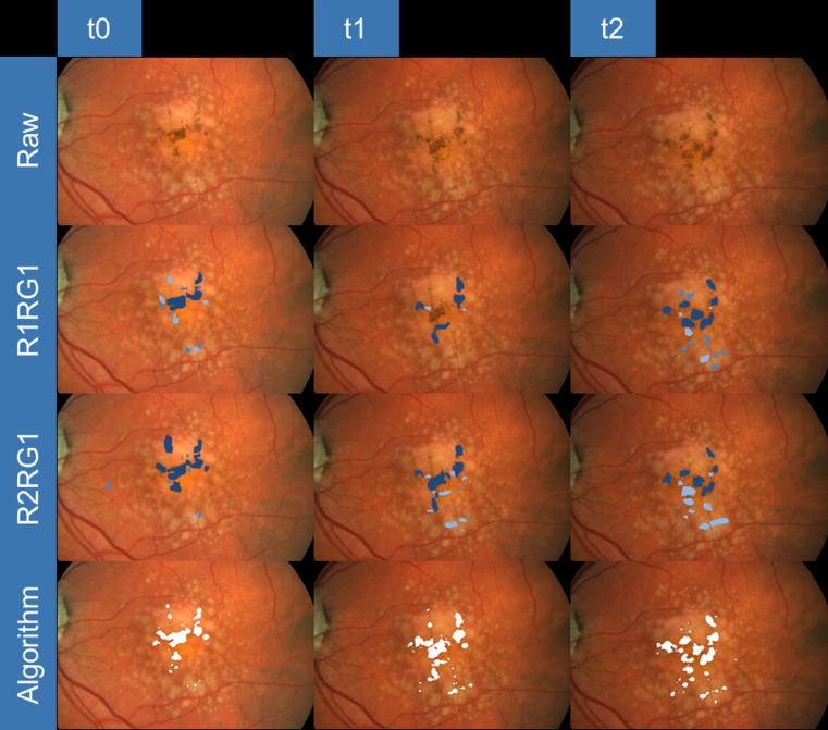
Color fundus (CFP) and autofluorescence (AF) photographs of 33 eyes with FHC of 28 AMD patients (mean age 71 years) from the prospective longitudinal natural history MODIAMD-study were included. Fully automated to semiautomated registration of baseline to corresponding follow-up images was evaluated. Following the manual circumscription of individual FHC (four different readings by two readers), a machine-learning algorithm was evaluated for automatic FHC detection.

RESULTS

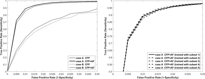
The overall pixel distance error for the semiautomated (CFP follow-up to CFP baseline: median 5.7; CFP to AF images from the same visit: median 6.5) was larger as compared for the automated image registration (4.5 and 5.7; < 0.001 and < 0.001). The total number of manually circumscribed objects and the corresponding total size varied between 637 to 1163 and 520,848 pixels to 924,860 pixels, respectively. Performance of the learning algorithms showed a sensitivity of 96% at a specificity level of 98% using information from both CFP and AF images and defining small areas of FHC ("speckle appearance") as "neutral."

CONCLUSIONS

FHC as a high-risk feature for progression of AMD to late stages can be automatically assessed at different time points with similar sensitivity and specificity as compared to manual outlining. Upon further development of the research prototype, this approach may be useful both in natural history and interventional large-scale studies for a more refined classification and risk assessment of eyes with intermediate AMD.

TRANSLATIONAL RELEVANCE

Automated FHC detection opens the door for a more refined and detailed classification and risk assessment of eyes with intermediate AMD in both natural history and future interventional studies.

摘要

目的

开发并评估一种软件工具,用于自动检测中度年龄相关性黄斑变性(AMD)患者眼睛中的局灶性色素沉着改变(FHC)。

方法

纳入了来自前瞻性纵向自然史MODIAMD研究的28例AMD患者(平均年龄71岁)的33只患有FHC的眼睛的彩色眼底(CFP)和自发荧光(AF)照片。评估了从基线到相应随访图像的全自动至半自动配准。在对个体FHC进行手动划定边界后(两名读者进行四次不同的读数),评估了一种机器学习算法用于自动检测FHC。

结果

与自动图像配准相比,半自动配准的总体像素距离误差更大(CFP随访至CFP基线:中位数5.7;同一就诊时CFP至AF图像:中位数6.5)(分别为4.5和5.7;<0.001和<0.001)。手动划定边界的对象总数和相应的总面积分别在637至1163个以及520,848像素至924,860像素之间变化。使用来自CFP和AF图像的信息并将FHC的小区域(“斑点外观”)定义为“中性”,学习算法的性能在特异性水平为98%时显示出96%的灵敏度。

结论

FHC作为AMD进展至晚期的高风险特征,可以在不同时间点进行自动评估,其灵敏度和特异性与手动勾勒相似。随着研究原型的进一步开发,这种方法可能在自然史和介入性大规模研究中都有用,以便对中度AMD患者的眼睛进行更精细的分类和风险评估。

转化相关性

自动检测FHC为在自然史和未来介入性研究中对中度AMD患者的眼睛进行更精细和详细的分类及风险评估打开了大门。

https://cdn.ncbi.nlm.nih.gov/pmc/blobs/dfd3/4782823/2b39d5b8ff15/i2164-2591-5-2-3-f04.jpg
https://cdn.ncbi.nlm.nih.gov/pmc/blobs/dfd3/4782823/c45c5ed43c5d/i2164-2591-5-2-3-f01.jpg
https://cdn.ncbi.nlm.nih.gov/pmc/blobs/dfd3/4782823/b6eddfd3ad63/i2164-2591-5-2-3-f02.jpg
https://cdn.ncbi.nlm.nih.gov/pmc/blobs/dfd3/4782823/f4360517eb90/i2164-2591-5-2-3-f03.jpg
https://cdn.ncbi.nlm.nih.gov/pmc/blobs/dfd3/4782823/2b39d5b8ff15/i2164-2591-5-2-3-f04.jpg

相似文献

1
Automated Retinal Image Analysis for Evaluation of Focal Hyperpigmentary Changes in Intermediate Age-Related Macular Degeneration.用于评估中度年龄相关性黄斑变性中局灶性色素沉着变化的自动视网膜图像分析
Transl Vis Sci Technol. 2016 Mar 4;5(2):3. doi: 10.1167/tvst.5.2.3. eCollection 2016 Mar.
2
Spatial correlation between hyperpigmentary changes on color fundus photography and hyperreflective foci on SDOCT in intermediate AMD.中度年龄相关性黄斑变性患者彩色眼底照片上的色素沉着改变与光谱域光学相干断层扫描上的高反射灶之间的空间相关性。
Invest Ophthalmol Vis Sci. 2012 Jul 9;53(8):4626-33. doi: 10.1167/iovs.12-9813.
3
ULTRAWIDEFIELD PSEUDOCOLOR RETINAL IMAGING VERSUS REAL-COLOR FUNDUS PHOTOGRAPHY FOR DETECTION OF INTRARETINAL PIGMENT MIGRATION IN AGE-RELATED MACULAR DEGENERATION.超宽视野伪彩视网膜成像与真彩眼底照相在年龄相关性黄斑变性中检测视网膜内色素迁移的比较。
Retina. 2021 Mar 1;41(3):563-571. doi: 10.1097/IAE.0000000000002886.
4
Predicting Progression of Age-Related Macular Degeneration Using OCT and Fundus Photography.使用 OCT 和眼底照相预测年龄相关性黄斑变性的进展。
Ophthalmol Retina. 2021 Feb;5(2):118-125. doi: 10.1016/j.oret.2020.06.026. Epub 2020 Jun 26.
5
Peripheral Retinal Changes Associated with Age-Related Macular Degeneration in the Age-Related Eye Disease Study 2: Age-Related Eye Disease Study 2 Report Number 12 by the Age-Related Eye Disease Study 2 Optos PEripheral RetinA (OPERA) Study Research Group.与年龄相关性黄斑变性相关的周边视网膜变化:年龄相关性眼病研究 2:年龄相关性眼病研究 2 报告编号 12,由年龄相关性眼病研究 2 Optos 周边视网膜 A(OPERA)研究小组提供。
Ophthalmology. 2017 Apr;124(4):479-487. doi: 10.1016/j.ophtha.2016.12.004. Epub 2017 Jan 12.
6
Age-related macular degeneration and risk factors for the development of choroidal neovascularisation in the fellow eye: a 3-year follow-up study.年龄相关性黄斑变性及对侧眼脉络膜新生血管发生的危险因素:一项 3 年随访研究。
Ophthalmologica. 2011;226(3):110-8. doi: 10.1159/000329473. Epub 2011 Aug 3.
7
Drusen regression is associated with local changes in fundus autofluorescence in intermediate age-related macular degeneration.玻璃膜疣消退与年龄相关性中间型黄斑变性的眼底自发荧光的局部变化有关。
Am J Ophthalmol. 2013 Sep;156(3):532-542.e1. doi: 10.1016/j.ajo.2013.04.031. Epub 2013 Jul 3.
8
Automatic drusen quantification and risk assessment of age-related macular degeneration on color fundus images.自动玻璃膜疣定量分析和年龄相关性黄斑变性的风险评估在眼底彩色图像上。
Invest Ophthalmol Vis Sci. 2013 Apr 30;54(4):3019-27. doi: 10.1167/iovs.12-11449.
9
Prognostic Value of Retinal Layers in Comparison with Other Risk Factors for Conversion of Intermediate Age-related Macular Degeneration.视网膜层与其他危险因素相比对中间型年龄相关性黄斑变性转归的预测价值。
Ophthalmol Retina. 2020 Jan;4(1):31-40. doi: 10.1016/j.oret.2019.08.003. Epub 2019 Aug 20.
10
Spectral-domain optical coherence tomography characteristics of intermediate age-related macular degeneration.光谱域光相干断层扫描在年龄相关性中间型黄斑变性中的特征。
Ophthalmology. 2013 Jan;120(1):140-50. doi: 10.1016/j.ophtha.2012.07.004. Epub 2012 Sep 8.

引用本文的文献

1
Deep Learning to Distinguish Edema Secondary to Retinal Vein Occlusion and Diabetic Macular Edema: A Multimodal Approach Using OCT and Infrared Imaging.深度学习用于区分视网膜静脉阻塞继发水肿和糖尿病性黄斑水肿:一种使用光学相干断层扫描(OCT)和红外成像的多模态方法。
J Clin Med. 2025 Feb 5;14(3):1008. doi: 10.3390/jcm14031008.
2
Artificial intelligence applications in ophthalmic optical coherence tomography: a 12-year bibliometric analysis.人工智能在眼科光学相干断层扫描中的应用:一项为期12年的文献计量分析。
Int J Ophthalmol. 2024 Dec 18;17(12):2295-2307. doi: 10.18240/ijo.2024.12.19. eCollection 2024.
3
Multi-label classification of retinal disease via a novel vision transformer model.

本文引用的文献

1
DEVELOPMENT OF INTRARETINAL CYSTOID LESIONS IN EYES WITH INTERMEDIATE AGE-RELATED MACULAR DEGENERATION.中年年龄相关性黄斑变性患者眼内视网膜囊肿样病变的发展
Retina. 2016 Aug;36(8):1548-56. doi: 10.1097/IAE.0000000000000951.
2
Reticular drusen in eyes with high-risk characteristics for progression to late-stage age-related macular degeneration.具有进展为晚期年龄相关性黄斑变性高风险特征的眼睛中的网状玻璃膜疣。
Br J Ophthalmol. 2015 Sep;99(9):1289-94. doi: 10.1136/bjophthalmol-2014-306535. Epub 2015 Mar 20.
3
Automatic identification of reticular pseudodrusen using multimodal retinal image analysis.
通过新型视觉Transformer模型进行视网膜疾病的多标签分类
Front Neurosci. 2024 Jan 8;17:1290803. doi: 10.3389/fnins.2023.1290803. eCollection 2023.
4
Longitudinal Changes of Retinal Structure in Molecularly Confirmed C1QTNF5 Patients With Late-Onset Retinal Degeneration.分子确诊的晚发性视网膜变性 C1QTNF5 患者的视网膜结构的纵向变化。
Transl Vis Sci Technol. 2023 Dec 1;12(12):14. doi: 10.1167/tvst.12.12.14.
5
Accurate drusen segmentation in optical coherence tomography via order-constrained regression of retinal layer heights.基于视网膜层高度有序约束回归的光学相干断层扫描精准 drusen 分割。
Sci Rep. 2023 May 19;13(1):8162. doi: 10.1038/s41598-023-35230-4.
6
The Development and Clinical Application of Innovative Optical Ophthalmic Imaging Techniques.创新光学眼科成像技术的发展与临床应用
Front Med (Lausanne). 2022 Jun 30;9:891369. doi: 10.3389/fmed.2022.891369. eCollection 2022.
7
From Data to Deployment: The Collaborative Community on Ophthalmic Imaging Roadmap for Artificial Intelligence in Age-Related Macular Degeneration.从数据到部署:眼科成像人工智能相关的年龄相关性黄斑变性协作社区路线图。
Ophthalmology. 2022 May;129(5):e43-e59. doi: 10.1016/j.ophtha.2022.01.002. Epub 2022 Jan 10.
8
Multimodal Retinal Image Analysis via Deep Learning for the Diagnosis of Intermediate Dry Age-Related Macular Degeneration: A Feasibility Study.基于深度学习的多模态视网膜图像分析用于诊断中度干性年龄相关性黄斑变性:一项可行性研究
J Ophthalmol. 2020 Jan 13;2020:7493419. doi: 10.1155/2020/7493419. eCollection 2020.
9
Deep learning-based detection and classification of geographic atrophy using a deep convolutional neural network classifier.使用深度卷积神经网络分类器基于深度学习的地理性萎缩检测与分类
Graefes Arch Clin Exp Ophthalmol. 2018 Nov;256(11):2053-2060. doi: 10.1007/s00417-018-4098-2. Epub 2018 Aug 8.
使用多模态视网膜图像分析自动识别网状假性玻璃膜疣
Invest Ophthalmol Vis Sci. 2015 Jan 8;56(1):633-9. doi: 10.1167/iovs.14-15019.
4
Optical coherence tomography-defined changes preceding the development of drusen-associated atrophy in age-related macular degeneration.光学相干断层扫描定义的在年龄相关性黄斑变性相关的 drusen 相关萎缩发展之前的变化。
Ophthalmology. 2014 Dec;121(12):2415-22. doi: 10.1016/j.ophtha.2014.06.034. Epub 2014 Aug 8.
5
Macular dystrophies mimicking age-related macular degeneration.黄斑营养不良类似于年龄相关性黄斑变性。
Prog Retin Eye Res. 2014 Mar;39:23-57. doi: 10.1016/j.preteyeres.2013.11.001. Epub 2013 Nov 28.
6
Automated drusen segmentation and quantification in SD-OCT images.SD-OCT 图像中的自动 drusen 分割和定量。
Med Image Anal. 2013 Dec;17(8):1058-72. doi: 10.1016/j.media.2013.06.003. Epub 2013 Jul 2.
7
Optical coherence tomography-based observation of the natural history of drusenoid lesion in eyes with dry age-related macular degeneration.基于光学相干断层扫描的干性年龄相关性黄斑变性患者玻璃膜疣病变自然史的观察。
Ophthalmology. 2013 Dec;120(12):2656-2665. doi: 10.1016/j.ophtha.2013.05.029. Epub 2013 Jul 4.
8
Automatic drusen quantification and risk assessment of age-related macular degeneration on color fundus images.自动玻璃膜疣定量分析和年龄相关性黄斑变性的风险评估在眼底彩色图像上。
Invest Ophthalmol Vis Sci. 2013 Apr 30;54(4):3019-27. doi: 10.1167/iovs.12-11449.
9
Progression of intermediate age-related macular degeneration with proliferation and inner retinal migration of hyperreflective foci.伴有高反射性病灶增殖和内视网膜迁移的中老年相关性黄斑变性进展。
Ophthalmology. 2013 May;120(5):1038-45. doi: 10.1016/j.ophtha.2012.10.018. Epub 2013 Jan 23.
10
Clinical classification of age-related macular degeneration.年龄相关性黄斑变性的临床分类。
Ophthalmology. 2013 Apr;120(4):844-51. doi: 10.1016/j.ophtha.2012.10.036. Epub 2013 Jan 16.