Teos L Y, Zheng C-Y, Liu X, Swaim W D, Goldsmith C M, Cotrim A P, Baum B J, Ambudkar I S
Secretory and Physiology Section, Molecular Physiology and Therapeutics Branch, NIDCR, NIH, Bethesda, MD, USA.
Translational Research Core, Molecular Physiology and Therapeutics Branch, NIDCR, NIH, Bethesda, MD, USA.
Gene Ther. 2016 Jul;23(7):572-9. doi: 10.1038/gt.2016.29. Epub 2016 Mar 11.
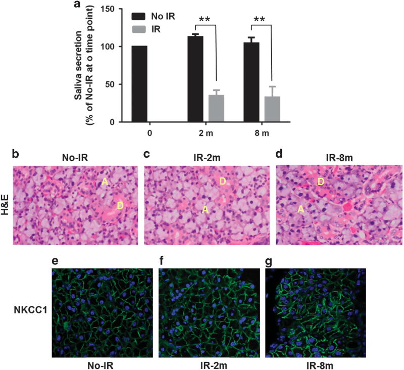
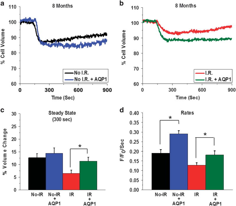
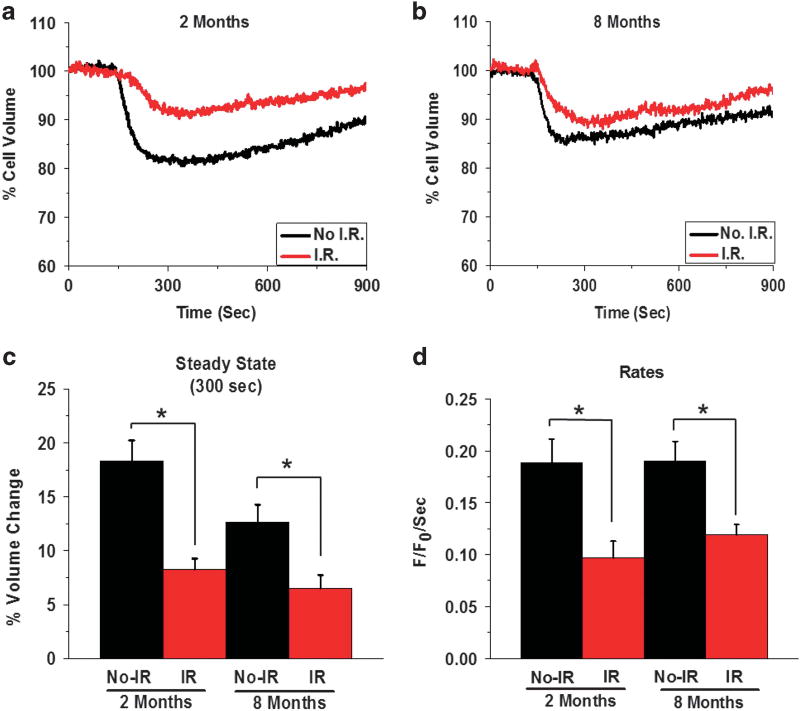
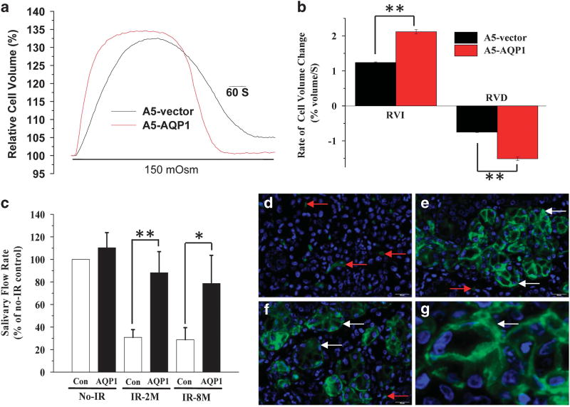
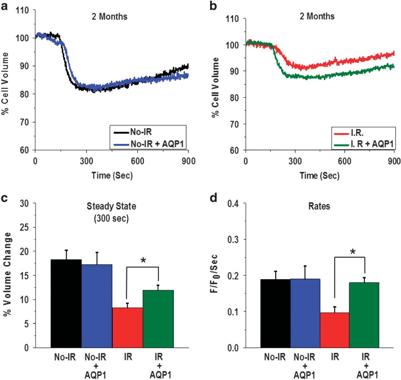
Head and neck irradiation (IR) during cancer treatment causes by-stander effects on the salivary glands leading to irreversible loss of saliva secretion. The mechanism underlying loss of fluid secretion is not understood and no adequate therapy is currently available. Delivery of an adenoviral vector encoding human aquaporin-1 (hAQP1) into the salivary glands of human subjects and animal models with radiation-induced salivary hypofunction leads to significant recovery of saliva secretion and symptomatic relief in subjects. To elucidate the mechanism underlying loss of salivary secretion and the basis for AdhAQP1-dependent recovery of salivary gland function we assessed submandibular gland function in control mice and mice 2 and 8 months after treatment with a single 15-Gy dose of IR (delivered to the salivary gland region). Salivary secretion and neurotransmitter-stimulated changes in acinar cell volume, an in vitro read-out for fluid secretion, were monitored. Consistent with the sustained 60% loss of fluid secretion following IR, a carbachol (CCh)-induced decrease in acinar cell volume from the glands of mice post IR was transient and attenuated as compared with that in cells from non-IR age-matched mice. The hAQP1 expression in non-IR mice induced no significant effect on salivary fluid secretion or CCh-stimulated cell volume changes, except in acinar cells from 8-month group where the initial rate of cell shrinkage was increased. Importantly, the expression of hAQP1 in the glands of mice post IR induced recovery of salivary fluid secretion and a volume decrease in acinar cells to levels similar to those in cells from non-IR mice. The initial rates of CCh-stimulated cell volume reduction in acinar cells from hAQP1-expressing glands post IR were similar to those from control cells. Altogether, the data suggest that expression of hAQP1 increases the water permeability of acinar cells, which underlies the recovery of fluid secretion in the salivary glands functionally compromised post IR.
癌症治疗期间对头颈部进行照射(IR)会对唾液腺产生旁观者效应,导致唾液分泌不可逆转地丧失。液体分泌丧失的潜在机制尚不清楚,目前也没有有效的治疗方法。将编码人水通道蛋白-1(hAQP1)的腺病毒载体导入患有放射性唾液腺功能减退的人类受试者和动物模型的唾液腺中,可使受试者的唾液分泌显著恢复并缓解症状。为了阐明唾液分泌丧失的潜在机制以及AdhAQP1依赖性唾液腺功能恢复的基础,我们评估了对照小鼠以及单次接受15 Gy剂量IR(照射唾液腺区域)后2个月和8个月的小鼠的下颌下腺功能。监测了唾液分泌以及腺泡细胞体积中神经递质刺激引起的变化,这是液体分泌的体外读数。与IR后持续60%的液体分泌丧失一致,与未接受IR的年龄匹配小鼠的细胞相比,IR后小鼠腺体中由卡巴胆碱(CCh)诱导的腺泡细胞体积减小是短暂的且减弱的。在未接受IR的小鼠中,hAQP1的表达对唾液分泌或CCh刺激的细胞体积变化没有显著影响,但在8个月组的腺泡细胞中,细胞初始收缩率有所增加。重要的是,IR后小鼠腺体中hAQP1的表达诱导唾液分泌恢复,腺泡细胞体积减小至与未接受IR小鼠细胞相似的水平。IR后hAQP1表达腺体的腺泡细胞中CCh刺激的细胞体积减小的初始速率与对照细胞相似。总之,数据表明hAQP1的表达增加了腺泡细胞的水通透性,这是IR后功能受损的唾液腺中液体分泌恢复的基础。