Wang Zhongqiu, Wu Wenzhong, Liu Yongkang, Wang Tianyao, Chen Xiao, Zhang Jianhua, Zhou Guoxing, Chen Rong
Department of Radiology, Affiliated Hospital of Nanjing University of Chinese Medicine, 155 Hanzhong Road, Nanjing 210029, Jiangsu, China.
Department of Acupuncture & Rehabilitation, Affiliated Hospital of Nanjing University of Chinese Medicine, 155 Hanzhong Road, Nanjing 210029, Jiangsu, China.
PLoS One. 2016 Mar 11;11(3):e0151489. doi: 10.1371/journal.pone.0151489. eCollection 2016.
Imaging studies of traumatic brain injury demonstrate that the cerebellum is often affected. We aim to examine fractional anisotropy alteration in acute-phase mild traumatic brain injury patients in cerebellum-related white matter tracts.
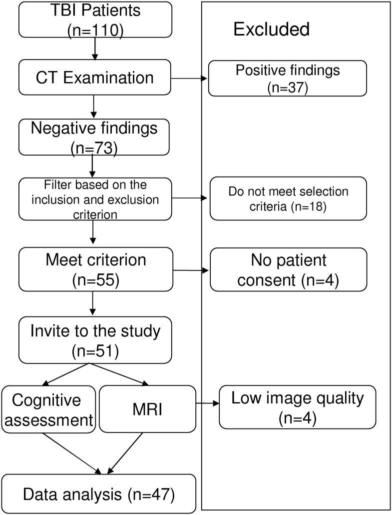
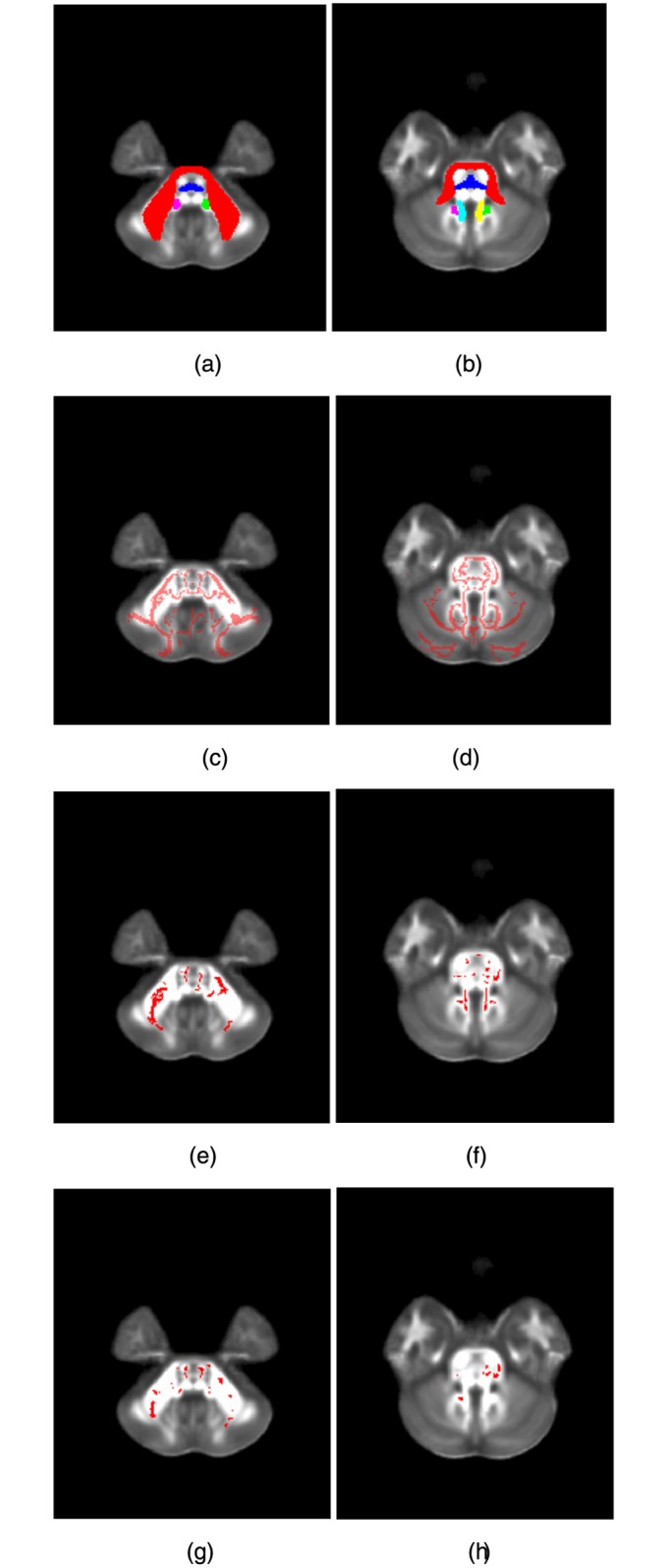
This prospective study included 47 mild traumatic brain injury patients in the acute stage and 37 controls. MR imaging and neurocognitive tests were performed in patients within 7 days of injury. White matter integrity was examined by using diffusion tensor imaging. We used three approaches, tract-based spatial statistics, graphical-model-based multivariate analysis, and region-of-interest analysis, to detect altered cerebellar white matter integrity in mild traumatic brain injury patients.
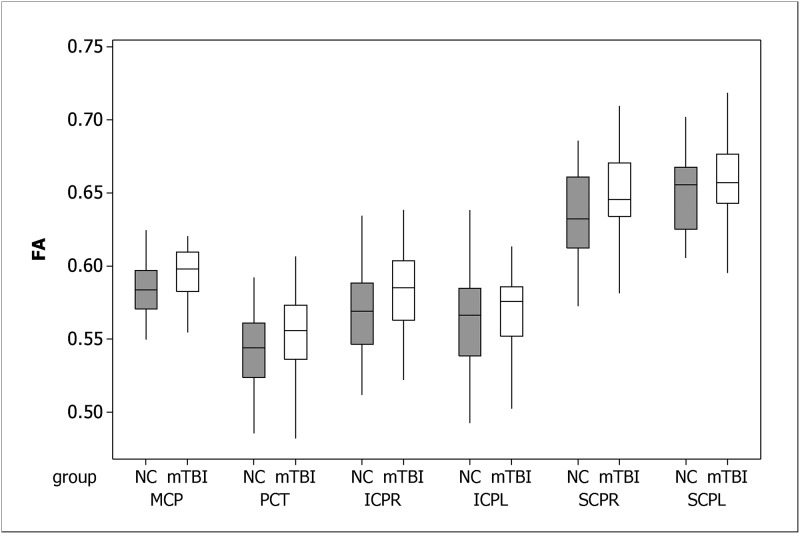
Results from three analysis methods were in accordance with each other, and suggested fractional anisotropy in the middle cerebellar peduncle and the pontine crossing tract was changed in the acute-phase mild traumatic brain injury patients, relative to controls (adjusted p-value < 0.05). Higher fractional anisotropy in the middle cerebellar peduncle was associated with worse performance in the fluid cognition composite (r = -0.289, p-value = 0.037).
Altered cerebellar fractional anisotropy in acute-phase mild traumatic brain injury patients is localized in specific regions and statistically associated with cognitive deficits detectable on neurocognitive testing.
创伤性脑损伤的影像学研究表明,小脑常受影响。我们旨在研究急性期轻度创伤性脑损伤患者小脑相关白质束的各向异性分数改变。
这项前瞻性研究纳入了47例急性期轻度创伤性脑损伤患者和37例对照者。在受伤7天内对患者进行了磁共振成像和神经认知测试。使用扩散张量成像检查白质完整性。我们采用了三种方法,即基于体素的空间统计学、基于图形模型的多变量分析和感兴趣区域分析,来检测轻度创伤性脑损伤患者小脑白质完整性的改变。
三种分析方法的结果相互一致,表明急性期轻度创伤性脑损伤患者相对于对照者,小脑中脚和脑桥交叉束的各向异性分数发生了改变(校正p值<0.05)。小脑中脚较高的各向异性分数与流体认知综合测试中较差的表现相关(r = -0.289,p值 = 0.037)。
急性期轻度创伤性脑损伤患者小脑各向异性分数的改变局限于特定区域,并且在神经认知测试中与可检测到的认知缺陷存在统计学关联。