• 文献检索
  • 文档翻译
  • 深度研究
  • 学术资讯
  • Suppr Zotero 插件Zotero 插件
  • 邀请有礼
  • 套餐&价格
  • 历史记录
应用&插件
Suppr Zotero 插件Zotero 插件浏览器插件Mac 客户端Windows 客户端微信小程序
定价
高级版会员购买积分包购买API积分包
服务
文献检索文档翻译深度研究API 文档MCP 服务
关于我们
关于 Suppr公司介绍联系我们用户协议隐私条款
关注我们

Suppr 超能文献

核心技术专利:CN118964589B侵权必究
粤ICP备2023148730 号-1Suppr @ 2026

文献检索

告别复杂PubMed语法,用中文像聊天一样搜索,搜遍4000万医学文献。AI智能推荐,让科研检索更轻松。

立即免费搜索

文件翻译

保留排版,准确专业,支持PDF/Word/PPT等文件格式,支持 12+语言互译。

免费翻译文档

深度研究

AI帮你快速写综述,25分钟生成高质量综述,智能提取关键信息,辅助科研写作。

立即免费体验

轻度创伤性脑损伤的磁共振成像和弥散张量成像研究进展综述。

A review of magnetic resonance imaging and diffusion tensor imaging findings in mild traumatic brain injury.

机构信息

Clinical Neuroscience Laboratory, Department of Psychiatry, VA Boston Healthcare System, Brockton, MA, USA.

出版信息

Brain Imaging Behav. 2012 Jun;6(2):137-92. doi: 10.1007/s11682-012-9156-5.

DOI:10.1007/s11682-012-9156-5
PMID:22438191
原文链接:https://pmc.ncbi.nlm.nih.gov/articles/PMC3803157/
Abstract

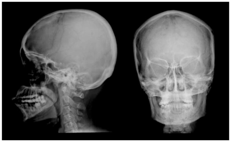
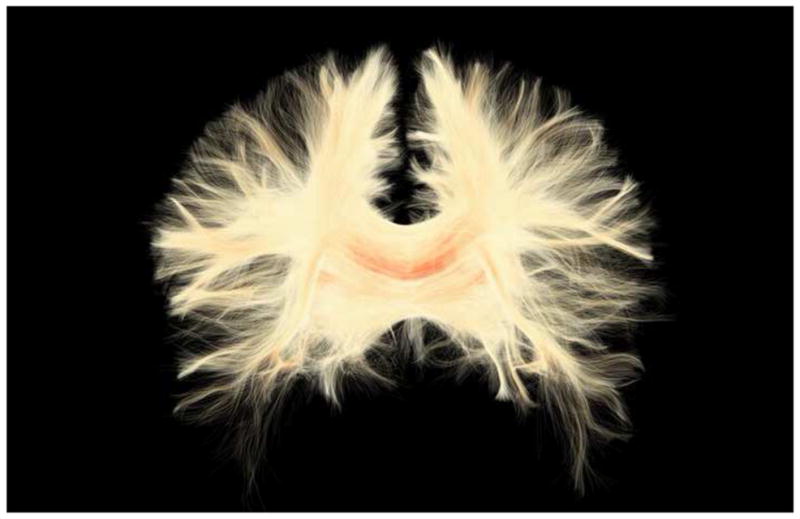
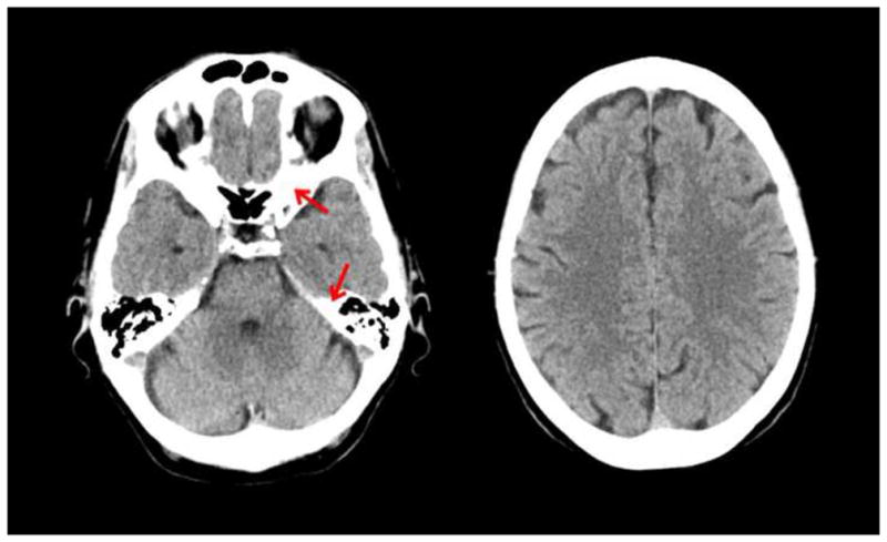
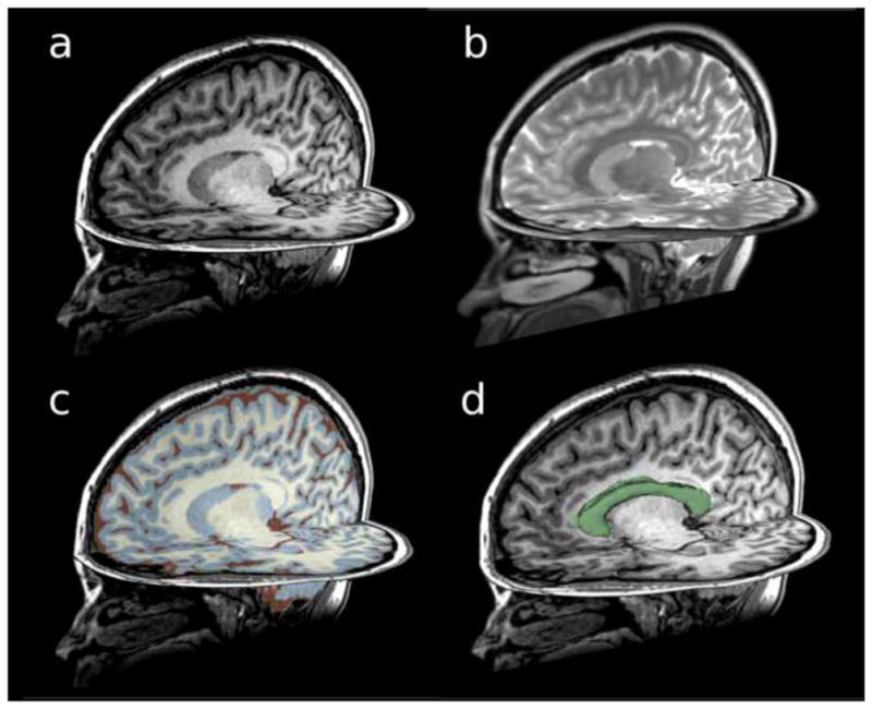
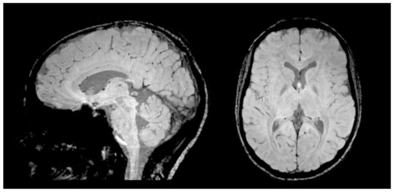
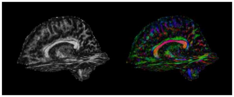
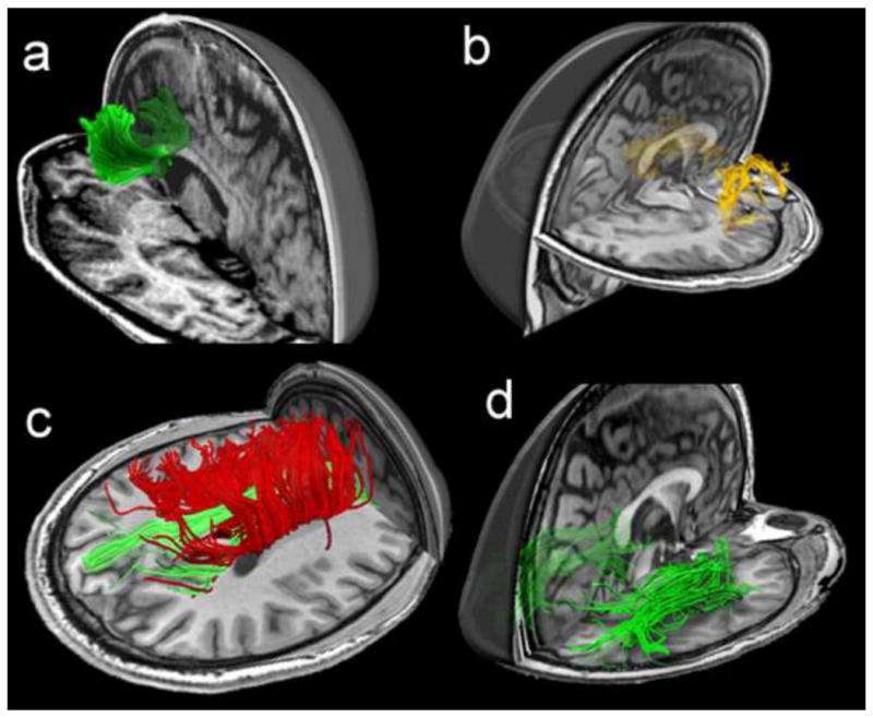
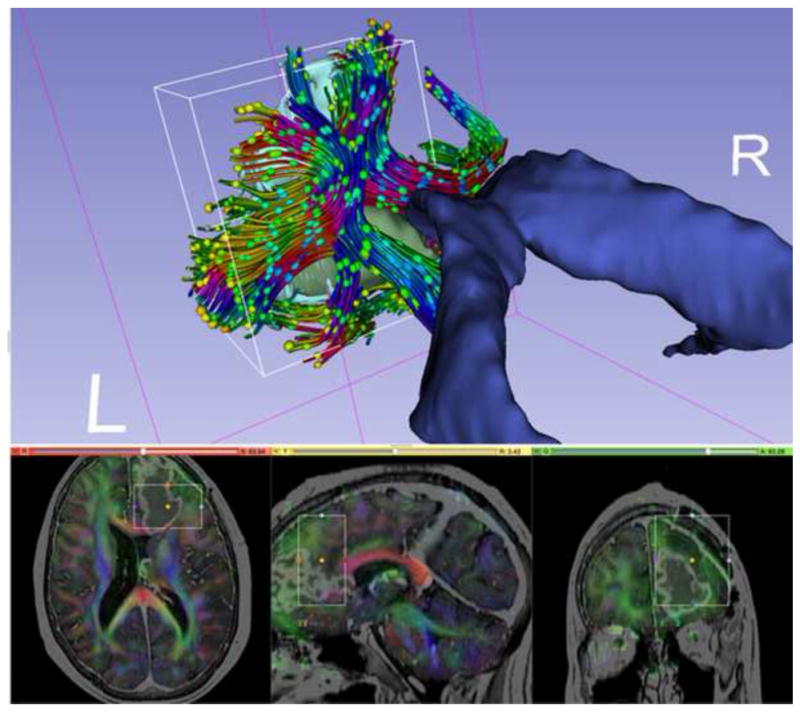
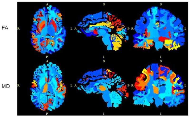
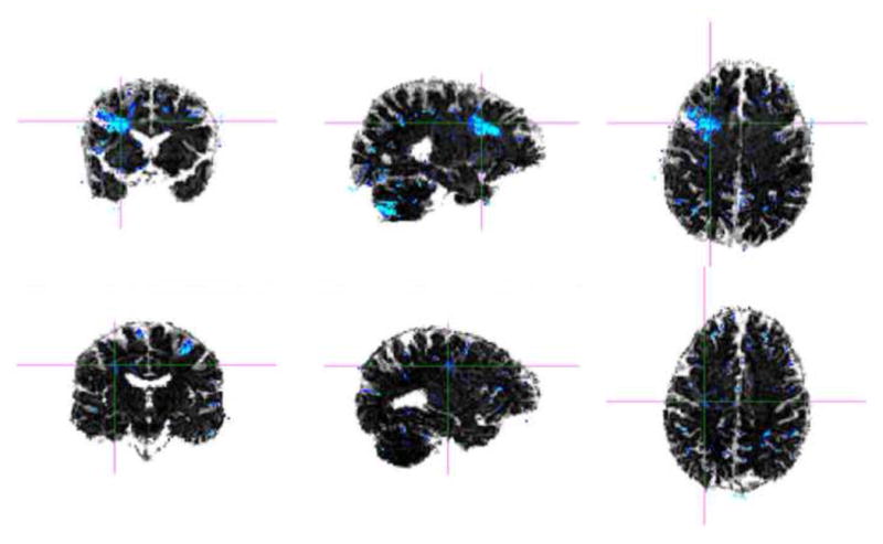
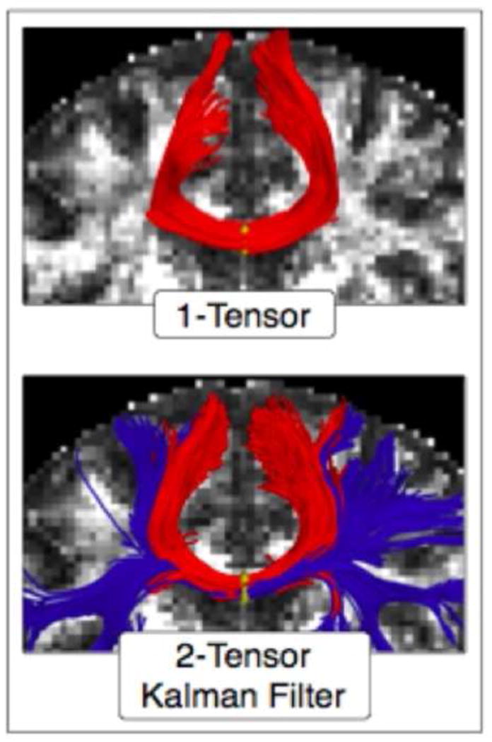
Mild traumatic brain injury (mTBI), also referred to as concussion, remains a controversial diagnosis because the brain often appears quite normal on conventional computed tomography (CT) and magnetic resonance imaging (MRI) scans. Such conventional tools, however, do not adequately depict brain injury in mTBI because they are not sensitive to detecting diffuse axonal injuries (DAI), also described as traumatic axonal injuries (TAI), the major brain injuries in mTBI. Furthermore, for the 15 to 30 % of those diagnosed with mTBI on the basis of cognitive and clinical symptoms, i.e., the "miserable minority," the cognitive and physical symptoms do not resolve following the first 3 months post-injury. Instead, they persist, and in some cases lead to long-term disability. The explanation given for these chronic symptoms, i.e., postconcussive syndrome, particularly in cases where there is no discernible radiological evidence for brain injury, has led some to posit a psychogenic origin. Such attributions are made all the easier since both posttraumatic stress disorder (PTSD) and depression are frequently co-morbid with mTBI. The challenge is thus to use neuroimaging tools that are sensitive to DAI/TAI, such as diffusion tensor imaging (DTI), in order to detect brain injuries in mTBI. Of note here, recent advances in neuroimaging techniques, such as DTI, make it possible to characterize better extant brain abnormalities in mTBI. These advances may lead to the development of biomarkers of injury, as well as to staging of reorganization and reversal of white matter changes following injury, and to the ability to track and to characterize changes in brain injury over time. Such tools will likely be used in future research to evaluate treatment efficacy, given their enhanced sensitivity to alterations in the brain. In this article we review the incidence of mTBI and the importance of characterizing this patient population using objective radiological measures. Evidence is presented for detecting brain abnormalities in mTBI based on studies that use advanced neuroimaging techniques. Taken together, these findings suggest that more sensitive neuroimaging tools improve the detection of brain abnormalities (i.e., diagnosis) in mTBI. These tools will likely also provide important information relevant to outcome (prognosis), as well as play an important role in longitudinal studies that are needed to understand the dynamic nature of brain injury in mTBI. Additionally, summary tables of MRI and DTI findings are included. We believe that the enhanced sensitivity of newer and more advanced neuroimaging techniques for identifying areas of brain damage in mTBI will be important for documenting the biological basis of postconcussive symptoms, which are likely associated with subtle brain alterations, alterations that have heretofore gone undetected due to the lack of sensitivity of earlier neuroimaging techniques. Nonetheless, it is noteworthy to point out that detecting brain abnormalities in mTBI does not mean that other disorders of a more psychogenic origin are not co-morbid with mTBI and equally important to treat. They arguably are. The controversy of psychogenic versus physiogenic, however, is not productive because the psychogenic view does not carefully consider the limitations of conventional neuroimaging techniques in detecting subtle brain injuries in mTBI, and the physiogenic view does not carefully consider the fact that PTSD and depression, and other co-morbid conditions, may be present in those suffering from mTBI. Finally, we end with a discussion of future directions in research that will lead to the improved care of patients diagnosed with mTBI.

摘要

轻度创伤性脑损伤(mTBI),也称为脑震荡,仍然是一个有争议的诊断,因为大脑在常规计算机断层扫描(CT)和磁共振成像(MRI)扫描中通常看起来非常正常。然而,这些常规工具并不能充分描绘 mTBI 中的脑损伤,因为它们对检测弥漫性轴索损伤(DAI)不够敏感,也称为创伤性轴索损伤(TAI),是 mTBI 中的主要脑损伤。此外,对于那些基于认知和临床症状被诊断为 mTBI 的人,即“痛苦的少数”,其中 15%至 30%的人在受伤后的头 3 个月内,认知和身体症状不会缓解。相反,它们会持续存在,在某些情况下会导致长期残疾。对于这些慢性症状,即脑震荡后综合征,尤其是在没有明显放射学证据的情况下,解释为心理起源,这导致一些人提出心理起源的说法。由于创伤后应激障碍(PTSD)和抑郁症经常与 mTBI 共病,因此这种归因更容易做出。因此,挑战在于使用对 DAI/TAI 敏感的神经影像学工具,如扩散张量成像(DTI),以检测 mTBI 中的脑损伤。这里值得注意的是,神经影像学技术的最新进展,如 DTI,使得更好地描述 mTBI 中现存的脑异常成为可能。这些进展可能导致损伤的生物标志物的发展,以及损伤后白质变化的重组和逆转的分期,以及追踪和描述脑损伤随时间的变化的能力。鉴于这些工具对大脑变化的敏感性更高,它们可能会在未来的研究中用于评估治疗效果。在本文中,我们回顾了 mTBI 的发生率,以及使用客观的放射学测量来描述这一患者群体的重要性。证据表明,基于使用先进神经影像学技术的研究,可以在 mTBI 中检测到脑异常。这些发现表明,更敏感的神经影像学工具可以提高 mTBI 中脑异常(即诊断)的检测能力。这些工具还可能提供与预后(预后)相关的重要信息,并在需要了解 mTBI 中脑损伤动态性质的纵向研究中发挥重要作用。此外,还包括 MRI 和 DTI 发现的摘要表。我们相信,更新和更先进的神经影像学技术在识别 mTBI 中脑损伤区域方面的更高敏感性,对于记录脑震荡后症状的生物学基础将是重要的,这些症状可能与微妙的大脑改变有关,这些改变由于早期神经影像学技术的敏感性不足而未被发现。然而,值得指出的是,在 mTBI 中检测到脑异常并不意味着与 mTBI 共病的其他更多心理起源的疾病没有被发现,并且同样需要治疗。它们可能是这样。然而,心理起源与生理起源之间的争议并没有产生什么效果,因为心理起源的观点没有仔细考虑常规神经影像学技术在检测 mTBI 中微妙脑损伤方面的局限性,而生理起源的观点也没有仔细考虑 PTSD 和抑郁症以及其他共病的存在,这些都可能存在于患有 mTBI 的人中。最后,我们以讨论未来的研究方向结束,这将导致对诊断为 mTBI 的患者的治疗得到改善。

https://cdn.ncbi.nlm.nih.gov/pmc/blobs/d2fa/3803157/664ba7fcc0b4/nihms520757f11a.jpg
https://cdn.ncbi.nlm.nih.gov/pmc/blobs/d2fa/3803157/2036fcd123e7/nihms520757f1.jpg
https://cdn.ncbi.nlm.nih.gov/pmc/blobs/d2fa/3803157/f32d1f2cbc4a/nihms520757f2.jpg
https://cdn.ncbi.nlm.nih.gov/pmc/blobs/d2fa/3803157/e840958520ee/nihms520757f3.jpg
https://cdn.ncbi.nlm.nih.gov/pmc/blobs/d2fa/3803157/9d775537f8bf/nihms520757f4.jpg
https://cdn.ncbi.nlm.nih.gov/pmc/blobs/d2fa/3803157/699b0b019976/nihms520757f5.jpg
https://cdn.ncbi.nlm.nih.gov/pmc/blobs/d2fa/3803157/7df6f1cece77/nihms520757f6.jpg
https://cdn.ncbi.nlm.nih.gov/pmc/blobs/d2fa/3803157/be586df3235d/nihms520757f7.jpg
https://cdn.ncbi.nlm.nih.gov/pmc/blobs/d2fa/3803157/f3668a26abd4/nihms520757f8.jpg
https://cdn.ncbi.nlm.nih.gov/pmc/blobs/d2fa/3803157/05af7ae2d857/nihms520757f9.jpg
https://cdn.ncbi.nlm.nih.gov/pmc/blobs/d2fa/3803157/1e69d077f3bb/nihms520757f10.jpg
https://cdn.ncbi.nlm.nih.gov/pmc/blobs/d2fa/3803157/664ba7fcc0b4/nihms520757f11a.jpg

相似文献

1
A review of magnetic resonance imaging and diffusion tensor imaging findings in mild traumatic brain injury.轻度创伤性脑损伤的磁共振成像和弥散张量成像研究进展综述。
Brain Imaging Behav. 2012 Jun;6(2):137-92. doi: 10.1007/s11682-012-9156-5.
2
Exploring Serum Biomarkers for Mild Traumatic Brain Injury探索轻度创伤性脑损伤的血清生物标志物
3
Neuropathology of Mild Traumatic Brain Injury: Correlation to Neurocognitive and Neurobehavioral Findings轻度创伤性脑损伤的神经病理学:与神经认知和神经行为结果的相关性
4
Magnetic Resonance Imaging Application in the Area of Mild and Acute Traumatic Brain Injury: Implications for Diagnostic Markers?磁共振成像在轻度和急性创伤性脑损伤领域的应用:对诊断标志物有何启示?
5
Mild traumatic brain injury: Is DTI ready for the courtroom?轻度创伤性脑损伤:弥散张量成像能用于法庭了吗?
Int J Law Psychiatry. 2018 Nov-Dec;61:50-63. doi: 10.1016/j.ijlp.2018.09.002. Epub 2018 Nov 1.
6
White matter abnormalities in mild traumatic brain injury with and without post-traumatic stress disorder: a subject-specific diffusion tensor imaging study.轻度创伤性脑损伤伴和不伴创伤后应激障碍的脑白质异常:一项基于个体的弥散张量成像研究。
Brain Imaging Behav. 2018 Jun;12(3):870-881. doi: 10.1007/s11682-017-9744-5.
7
The role of biomarkers and MEG-based imaging markers in the diagnosis of post-traumatic stress disorder and blast-induced mild traumatic brain injury.生物标志物和基于脑磁图的成像标志物在创伤后应激障碍和爆炸所致轻度创伤性脑损伤诊断中的作用。
Psychoneuroendocrinology. 2016 Jan;63:398-409. doi: 10.1016/j.psyneuen.2015.02.008. Epub 2015 Feb 23.
8
Susceptibility Weighted Imaging and White Matter Abnormality Findings in Service Members With Persistent Cognitive Symptoms Following Mild Traumatic Brain Injury.轻度创伤性脑损伤后持续存在认知症状的军人的磁敏感加权成像和白质异常表现
Mil Med. 2017 Mar;182(3):e1651-e1658. doi: 10.7205/MILMED-D-16-00132.
9
The nature of white matter abnormalities in blast-related mild traumatic brain injury.爆炸相关轻度创伤性脑损伤中白质异常的性质。
Neuroimage Clin. 2015 Apr 9;8:148-56. doi: 10.1016/j.nicl.2015.04.001. eCollection 2015.
10
Neuroimaging biomarkers in mild traumatic brain injury (mTBI).轻度创伤性脑损伤(mTBI)的神经影像学生物标志物。
Neuropsychol Rev. 2013 Sep;23(3):169-209. doi: 10.1007/s11065-013-9237-2. Epub 2013 Aug 24.

引用本文的文献

1
Abnormal brain morphology and morphological brain network in adults with acute mild traumatic brain injury.急性轻度创伤性脑损伤成人的脑形态异常及脑形态网络
Neuroreport. 2025 Oct 1;36(14):777-785. doi: 10.1097/WNR.0000000000002186. Epub 2025 Jul 21.
2
Early cerebrospinal fluid elevations of pTau-217 in severe traumatic brain injury subjects.重度创伤性脑损伤患者脑脊液中pTau-217早期升高。
Front Neurol. 2025 Jul 30;16:1632679. doi: 10.3389/fneur.2025.1632679. eCollection 2025.
3
Bundle-specific tractography approach for identifying white matter microstructural changes following traumatic brain injury in rats: An EpiBioS4Rx study.

本文引用的文献

1
Diffusion tensor imaging, structural connectivity, and schizophrenia.扩散张量成像、结构连接性与精神分裂症。
Schizophr Res Treatment. 2011;2011:709523. doi: 10.1155/2011/709523. Epub 2011 Jul 17.
2
Diffusion tensor imaging studies on corticospinal tract injury following traumatic brain injury: a review.弥散张量成像研究颅脑损伤后皮质脊髓束损伤: 综述。
NeuroRehabilitation. 2011;29(4):339-45. doi: 10.3233/NRE-2011-0710.
3
Subject-specific changes in brain white matter on diffusion tensor imaging after sports-related concussion.
用于识别大鼠创伤性脑损伤后白质微观结构变化的束特异性纤维束成像方法:一项EpiBioS4Rx研究。
Imaging Neurosci (Camb). 2025 Feb 18;3. doi: 10.1162/imag_a_00471. eCollection 2025.
4
Identifying key physiological and clinical factors for traumatic brain injury patient management using network analysis and machine learning.使用网络分析和机器学习识别创伤性脑损伤患者管理的关键生理和临床因素。
PLoS One. 2025 Jul 28;20(7):e0328870. doi: 10.1371/journal.pone.0328870. eCollection 2025.
5
Fiber tract integrity in patients with brain injury and chronic health symptoms.脑损伤和慢性健康症状患者的纤维束完整性
Neuroimage Rep. 2021 Sep 13;1(4):100047. doi: 10.1016/j.ynirp.2021.100047. eCollection 2021 Dec.
6
Assessment of White Matter Changes Using Quantitative T1ρ Mapping in an Open-Field Low-Intensity Blast Mouse Model of Mild Traumatic Brain Injury (mTBI).在轻度创伤性脑损伤(mTBI)的开放场低强度爆炸小鼠模型中,使用定量T1ρ成像评估白质变化。
Int J Mol Sci. 2025 Jun 6;26(12):5431. doi: 10.3390/ijms26125431.
7
Biomarkers of traumatic brain injury: narrative review and future prospects in neurointensive care.创伤性脑损伤的生物标志物:叙述性综述及神经重症监护的未来前景
Front Med (Lausanne). 2025 Jun 3;12:1539159. doi: 10.3389/fmed.2025.1539159. eCollection 2025.
8
Aggressive intracranial meningioma associated with multiple sclerosis: A case report and literature review.侵袭性颅内脑膜瘤合并多发性硬化:一例报告及文献复习
Med Int (Lond). 2025 May 21;5(4):44. doi: 10.3892/mi.2025.243. eCollection 2025 Jul-Aug.
9
White Matter Microstructural Correlates of Swallowing Biomechanics: An Exploratory Pilot Study in Healthy Young Adults.吞咽生物力学的白质微观结构关联:一项针对健康年轻成年人的探索性初步研究。
Dysphagia. 2025 May 27. doi: 10.1007/s00455-025-10841-3.
10
Revisiting the critical roles of reactive microglia in traumatic brain injury.重新审视反应性小胶质细胞在创伤性脑损伤中的关键作用。
Int J Surg. 2025 Jun 1;111(6):3942-3978. doi: 10.1097/JS9.0000000000002420. Epub 2025 May 12.
运动相关性脑震荡后弥散张量成像脑白质的特定部位变化。
Magn Reson Imaging. 2012 Feb;30(2):171-80. doi: 10.1016/j.mri.2011.10.001. Epub 2011 Nov 12.
4
Diffuse and spatially variable white matter disruptions are associated with blast-related mild traumatic brain injury.弥漫性和空间变异性的脑白质损伤与爆炸相关的轻度创伤性脑损伤有关。
Neuroimage. 2012 Feb 1;59(3):2017-24. doi: 10.1016/j.neuroimage.2011.10.050. Epub 2011 Oct 20.
5
Long-term consequences of repetitive brain trauma: chronic traumatic encephalopathy.重复性脑创伤的长期后果:慢性创伤性脑病。
PM R. 2011 Oct;3(10 Suppl 2):S460-7. doi: 10.1016/j.pmrj.2011.08.008.
6
Maximum principal strain and strain rate associated with concussion diagnosis correlates with changes in corpus callosum white matter indices.最大主应变和应变率与脑震荡诊断相关,与胼胝体白质指数的变化相关。
Ann Biomed Eng. 2012 Jan;40(1):127-40. doi: 10.1007/s10439-011-0402-6. Epub 2011 Oct 13.
7
How and how not to correct for CSF-contamination in diffusion MRI.如何以及如何避免在扩散 MRI 中纠正 CSF 污染。
Neuroimage. 2012 Jan 16;59(2):1394-403. doi: 10.1016/j.neuroimage.2011.08.043. Epub 2011 Sep 5.
8
Acute and chronic changes in diffusivity measures after sports concussion.运动性脑震荡后扩散测量的急性和慢性变化。
J Neurotrauma. 2011 Oct;28(10):2049-59. doi: 10.1089/neu.2011.1836. Epub 2011 Oct 4.
9
Diffusion kurtosis as an in vivo imaging marker for reactive astrogliosis in traumatic brain injury.扩散峰度成像作为创伤性脑损伤中反应性星形胶质增生的活体成像标志物。
Neuroimage. 2012 Jan 2;59(1):467-77. doi: 10.1016/j.neuroimage.2011.07.050. Epub 2011 Jul 30.
10
Comparison of acute and chronic traumatic brain injury using semi-automatic multimodal segmentation of MR volumes.使用磁共振容积半自动多模态分割比较急性和慢性创伤性脑损伤。
J Neurotrauma. 2011 Nov;28(11):2287-306. doi: 10.1089/neu.2011.1920. Epub 2011 Sep 21.