Schimmer Ralph C, Urner Martin, Voigtsberger Stefanie, Booy Christa, Roth Z'Graggen Birgit, Beck-Schimmer Beatrice, Schläpfer Martin
Department of Surgery, University Hospital Zurich, Zurich, Switzerland.
Institute of Anesthesiology, University Hospital Zurich, Zurich, Switzerland.
PLoS One. 2016 Mar 17;11(3):e0151903. doi: 10.1371/journal.pone.0151903. eCollection 2016.
Tissue hypoperfusion and inflammation in sepsis can lead to organ failure including kidney and liver. In sepsis, mortality of acute kidney injury increases by more than 50%. Which type of volume replacement should be used is still an ongoing debate. We investigated the effect of different volume strategies on inflammatory mediators in kidney and liver in an early sepsis model.
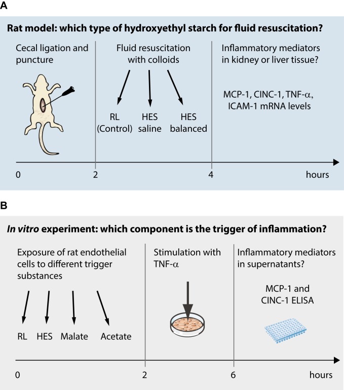
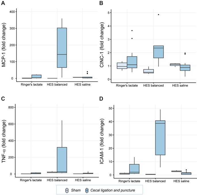
Adult male Wistar rats were subjected to sepsis by cecal ligation and puncture (CLP) and assigned to three fluid replenishment groups. Animals received 30mL/kg of Ringer's lactate (RL) for 2h, thereafter RL (75mL/kg), hydroxyethyl starch (HES) balanced (25mL/kg), containing malate and acetate, or HES saline (25mL/kg) for another 2h. Kidney and liver tissue was assessed for inflammation. In vitro rat endothelial cells were exposed to RL, HES balanced or HES saline for 2h, followed by stimulation with tumor necrosis factor-α (TNF-α) for another 4h. Alternatively, cells were exposed to malate, acetate or a mixture of malate and acetate, reflecting the according concentration of these substances in HES balanced. Pro-inflammatory cytokines were determined in cell supernatants.
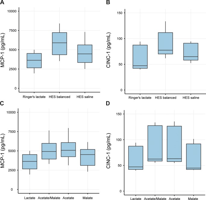
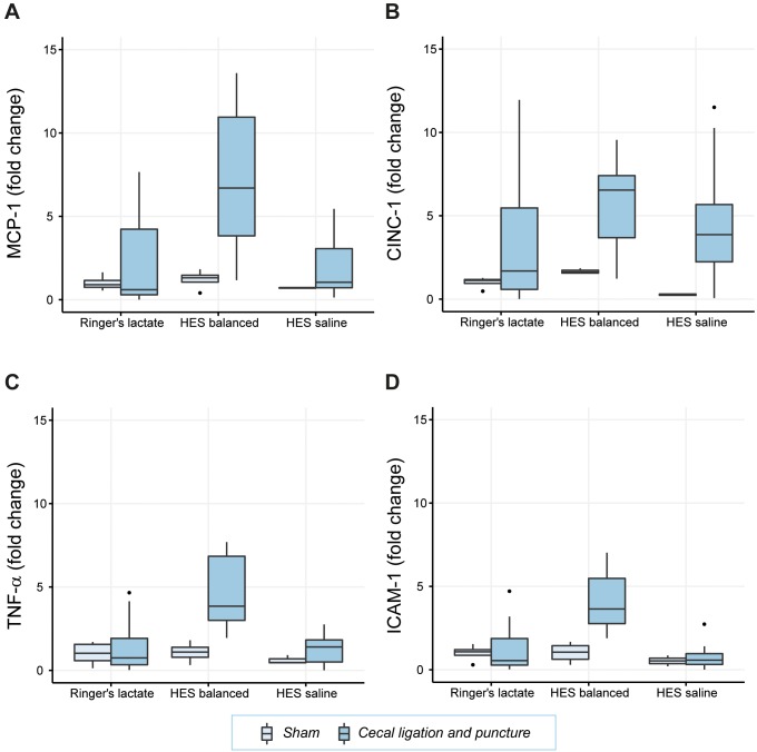
Cytokine mRNA in kidney and liver was increased in CLP animals treated with HES balanced compared to RL, but not after application of HES saline. MCP-1 was 3.5fold (95% CI: 1.3, 5.6) (p<0.01) and TNF-α 2.3fold (95% CI: 1.2, 3.3) (p<0.001) upregulated in the kidney. Corresponding results were seen in liver tissue. TNF-α-stimulated endothelial cells co-exposed to RL expressed 3529±1040pg/mL MCP-1 and 59±23pg/mL CINC-1 protein. These cytokines increased by 2358pg/mL (95% CI: 1511, 3204) (p<0.001) and 29pg/ml (95% CI: 14, 45) (p<0.01) respectively when exposed to HES balanced instead. However, no further upregulation was observed with HES saline. PBS supplemented with acetate increased MCP-1 by 1325pg/mL (95% CI: 741, 1909) (p<0.001) and CINC-1 by 24pg/mL (95% CI: 9, 38) (p<0.01) compared to RL. Malate as well as HES saline did not affect cytokine expression.
We identified HES balanced and specifically its component acetate as pro-inflammatory factor. How important this additional inflammatory burden on kidney and liver function is contributing to the sepsis-associated inflammatory burden in early sepsis needs further evaluation.
脓毒症中的组织灌注不足和炎症可导致包括肾和肝在内的器官衰竭。在脓毒症中,急性肾损伤的死亡率增加超过50%。应使用哪种类型的液体复苏仍在持续争论中。我们在早期脓毒症模型中研究了不同液体策略对肾和肝中炎症介质的影响。
成年雄性Wistar大鼠通过盲肠结扎和穿刺(CLP)诱导脓毒症,并分为三个液体补充组。动物先接受30mL/kg乳酸林格液(RL)输注2小时,之后分别接受RL(75mL/kg)、平衡型羟乙基淀粉(HES)(含苹果酸和醋酸盐,25mL/kg)或HES生理盐水(25mL/kg)再输注2小时。对肾和肝组织进行炎症评估。体外将大鼠内皮细胞暴露于RL、平衡型HES或HES生理盐水2小时,随后再用肿瘤坏死因子-α(TNF-α)刺激4小时。或者,将细胞暴露于苹果酸、醋酸盐或苹果酸与醋酸盐的混合物中,这些物质反映了平衡型HES中这些物质的相应浓度。测定细胞上清液中的促炎细胞因子。
与RL组相比,接受平衡型HES治疗的CLP动物肾和肝中的细胞因子mRNA增加,但应用HES生理盐水后未增加。肾中单核细胞趋化蛋白-1(MCP-1)上调3.5倍(95%置信区间:1.3,5.6)(p<0.01),TNF-α上调2.3倍(95%置信区间:1.2,3.3)(p<0.001)。肝组织中也观察到相应结果。与RL共同暴露于TNF-α刺激的内皮细胞表达3529±1040pg/mL的MCP-1和59±23pg/mL的CINC-1蛋白。当改为暴露于平衡型HES时,这些细胞因子分别增加了2358pg/mL(95%置信区间:1511,3204)(p<0.001)和29pg/ml(95%置信区间:14,45)(p<0.01)。然而,HES生理盐水未观察到进一步上调。与RL相比,补充醋酸盐的磷酸盐缓冲液使MCP-1增加1325pg/mL(95%置信区间:741,1909)(p<0.001),使CINC-1增加24pg/mL(95%置信区间:9,38)(p<0.01)。苹果酸以及HES生理盐水均不影响细胞因子表达。
我们确定平衡型HES及其特定成分醋酸盐为促炎因子。这种对肾和肝功能的额外炎症负担在早期脓毒症中对脓毒症相关炎症负担的影响程度需要进一步评估。