Aman M Javad
Integrated BioTherapeutics, Inc., Gaithersburg, Maryland, USA
mBio. 2016 Mar 22;7(2):e00346. doi: 10.1128/mBio.00346-16.
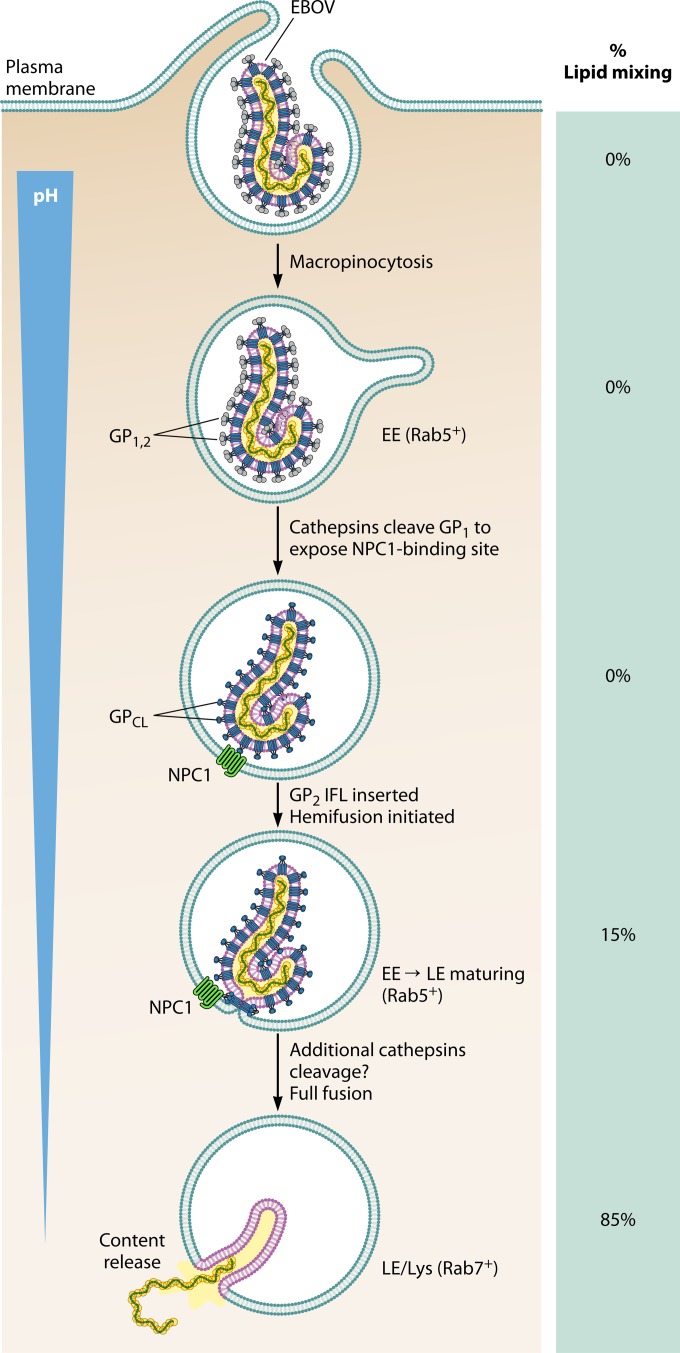
During virus entry, the surface glycoprotein of Ebola virus (EBOV) undergoes a complex set of transformations within the endosomal network. Tools to study EBOV entry have been limited to static immunofluorescence or biochemical and functional analysis. In a recent article inmBio, Spence et al. reported a novel, live-cell-imaging method that tracks this transformational journey of EBOV in real time [J. S. Spence, T. B. Krause, E. Mittler, R. K. Jangra, and K. Chandran, mBio 7(1):e01857-15, 2016, http://dx.doi.org/10.1128/mBio.01857-15]. The assay validates known mechanisms of EBOV entry and sheds light on some novel intricacies. Direct evidence supports the hypothesis that fusion is a rare event that starts in maturing early endosomes, is completed in late endosomes, and occurs entirely in Niemann-Pick C1 (NPC1)-positive (NPC1(+)) compartments. The study demonstrated that lipid mixing and productive fusion are temporally decoupled, with different energetic barriers and a protease-dependent step between the two events. Analysis of the mechanism of action of an important class of EBOV neutralizing antibodies, such as KZ52 and ZMapp, provides direct evidence that these antibodies act by inhibiting the membrane fusion.
在病毒进入过程中,埃博拉病毒(EBOV)的表面糖蛋白在内体网络内经历一系列复杂的转变。研究EBOV进入的工具仅限于静态免疫荧光或生化及功能分析。在最近发表于《mBio》的一篇文章中,斯彭斯等人报道了一种新颖的活细胞成像方法,该方法可实时追踪EBOV的这一转变过程[J. S. 斯彭斯、T. B. 克劳斯、E. 米特勒、R. K. 詹格拉和K. 钱德兰,《mBio》7(1):e01857 - 15,2016,http://dx.doi.org/10.1128/mBio.01857 - 15]。该检测方法验证了已知的EBOV进入机制,并揭示了一些新的复杂情况。直接证据支持了这样的假说,即融合是一种罕见事件,始于早期内体成熟过程,在晚期内体中完成,且完全发生在尼曼 - 匹克C1(NPC1)阳性(NPC1(+))区室中。该研究表明,脂质混合和有效融合在时间上是解耦的,两者之间存在不同的能量屏障以及一个蛋白酶依赖性步骤。对一类重要的EBOV中和抗体(如KZ52和ZMapp)的作用机制分析提供了直接证据,表明这些抗体通过抑制膜融合发挥作用。