Sayers Sophie R, Reimann Frank, Gribble Fiona M, Parker Helen, Zac-Varghese Sagen, Bloom Stephen R, Foretz Marc, Viollet Benoit, Rutter Guy A
Department of Cell Biology and Functional Genomics, Imperial College London, London, W12 ONN, United Kingdom.
Wellcome Trust - MRC Institute of Metabolic Science, University of Cambridge, Hills Road, Cambridge, CB2 0QQ, United Kingdom.
PLoS One. 2016 Mar 24;11(3):e0149549. doi: 10.1371/journal.pone.0149549. eCollection 2016.
Enteroendocrine L-cells synthesise and release the gut hormone glucagon-like peptide-1 (GLP-1) in response to food transit. Deletion of the tumour suppressor kinase LKB1 from proglucagon-expressing cells leads to the generation of intestinal polyps but no change in circulating GLP-1 levels. Here, we explore the role of the downstream kinase AMP-activated protein kinase (AMPK) in these cells.
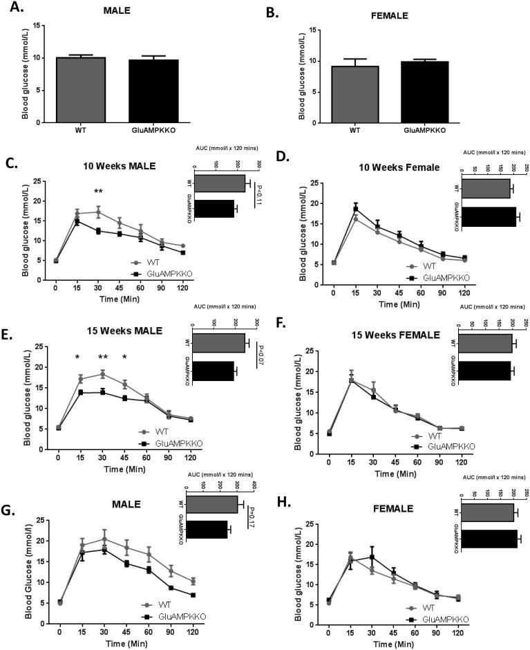
Loss of AMPK from proglucagon-expressing cells was achieved using a preproglucagon promoter-driven Cre (iGluCre) to catalyse recombination of floxed alleles of AMPKα1 and α2. Oral and intraperitoneal glucose tolerance were measured using standard protocols. L-cell mass was measured by immunocytochemistry. Hormone and peptide levels were measured by electrochemical-based luminescence detection or radioimmunoassay.
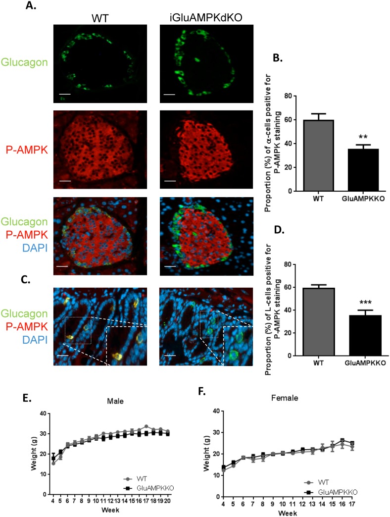
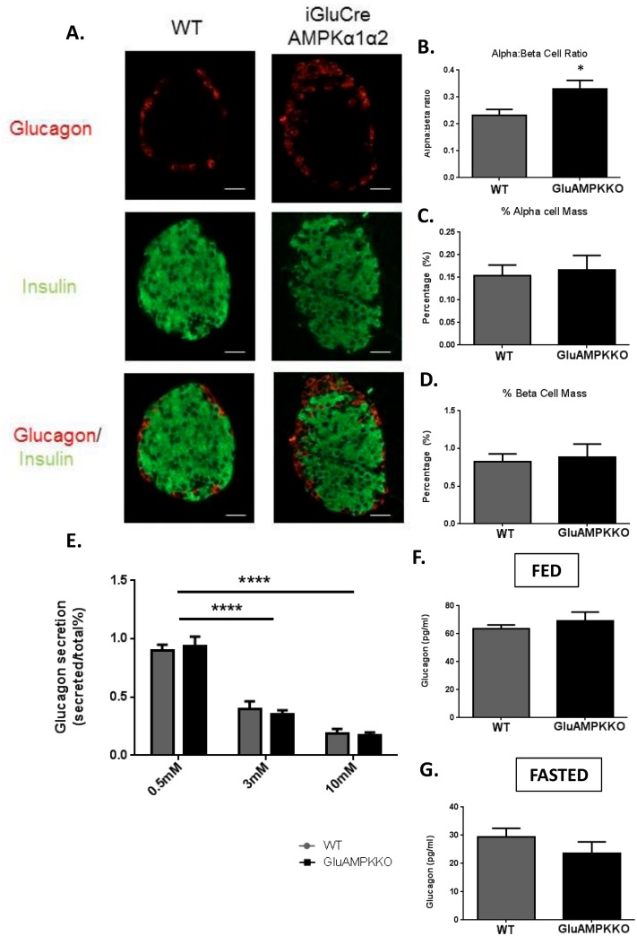
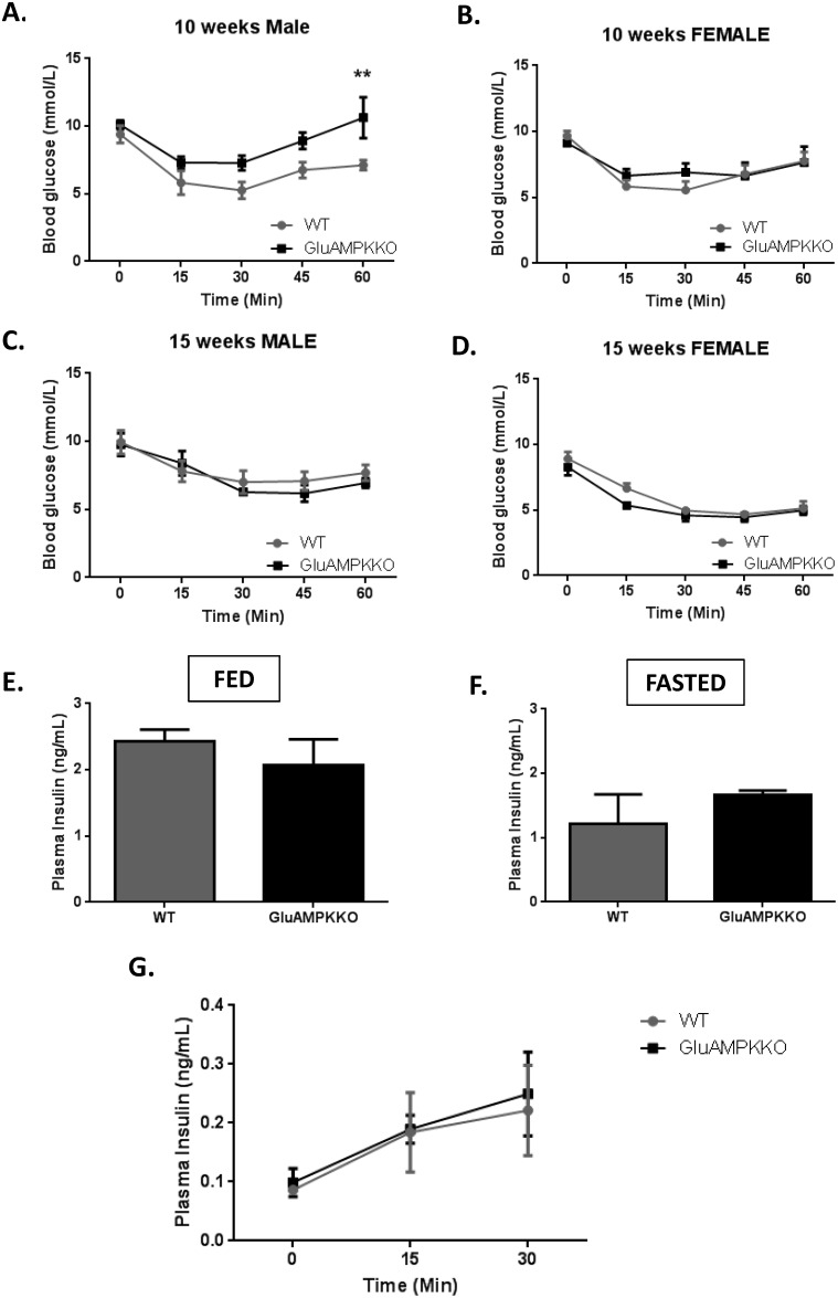
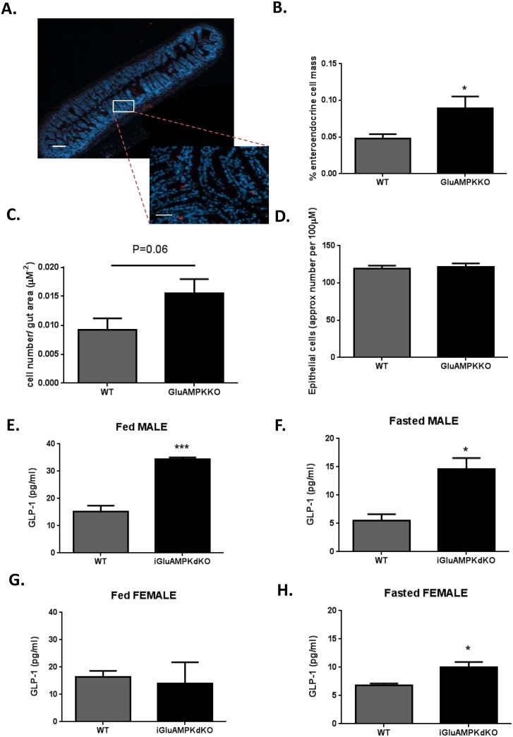
Recombination with iGluCre led to efficient deletion of AMPK from intestinal L- and pancreatic alpha-cells. In contrast to mice rendered null for LKB1 using the same strategy, mice deleted for AMPK displayed an increase (WT: 0.05 ± 0.01, KO: 0.09±0.02%, p<0.01) in L-cell mass and elevated plasma fasting (WT: 5.62 ± 0.800 pg/ml, KO: 14.5 ± 1.870, p<0.01) and fed (WT: 15.7 ± 1.48pg/ml, KO: 22.0 ± 6.62, p<0.01) GLP-1 levels. Oral, but not intraperitoneal, glucose tolerance was significantly improved by AMPK deletion, whilst insulin and glucagon levels were unchanged despite an increase in alpha to beta cell ratio (WT: 0.23 ± 0.02, KO: 0.33 ± 0.03, p<0.01).
AMPK restricts L-cell growth and GLP-1 secretion to suppress glucose tolerance. Targeted inhibition of AMPK in L-cells may thus provide a new therapeutic strategy in some forms of type 2 diabetes.
肠内分泌L细胞在食物通过时合成并释放肠道激素胰高血糖素样肽-1(GLP-1)。从表达胰高血糖素原的细胞中删除肿瘤抑制激酶LKB1会导致肠息肉的产生,但循环中的GLP-1水平没有变化。在此,我们探究下游激酶AMP激活的蛋白激酶(AMPK)在这些细胞中的作用。
利用前胰高血糖素启动子驱动的Cre(iGluCre)来催化AMPKα1和α2的floxed等位基因重组,从而实现从表达胰高血糖素原的细胞中删除AMPK。使用标准方案测量口服和腹腔内葡萄糖耐量。通过免疫细胞化学测量L细胞质量。通过基于电化学的发光检测或放射免疫测定法测量激素和肽水平。
与iGluCre重组导致肠道L细胞和胰腺α细胞中的AMPK有效缺失。与使用相同策略使LKB1缺失的小鼠不同,AMPK缺失的小鼠L细胞质量增加(野生型:0.05±0.01,敲除型:0.09±0.02%,p<0.01),血浆空腹(野生型:5.62±0.800 pg/ml,敲除型:14.5±1.870,p<0.01)和进食后(野生型:15.7±1.48 pg/ml,敲除型:22.0±6.62,p<0.01)的GLP-1水平升高。AMPK缺失显著改善了口服葡萄糖耐量,但腹腔内葡萄糖耐量未改善,尽管α细胞与β细胞的比例增加(野生型:0.23±0.02,敲除型:0.33±0.03,p<0.01),但胰岛素和胰高血糖素水平未改变。
AMPK限制L细胞生长和GLP-1分泌以抑制葡萄糖耐量。因此,对L细胞中AMPK的靶向抑制可能为某些形式的2型糖尿病提供一种新的治疗策略。