Thombare Ketan, Ntika Stelia, Wang Xuan, Krizhanovskii Camilla
Södertälje Hospital, Department of Internal Medicine, Södertälje, Sweden.
Karolinska Institute, Department of Molecular Medicine and Surgery, Stockholm, Sweden.
PLoS One. 2017 May 16;12(5):e0177605. doi: 10.1371/journal.pone.0177605. eCollection 2017.
Fatty acids acutely stimulate GLP-1 secretion from L-cells in vivo. However, a high fat diet has been shown to reduce the density of L-cells in the mouse intestine and a positive correlation has been indicated between L-cell number and GLP-1 secretion. Thus, the mechanism of fatty acid-stimulated GLP-1 secretion, potential effects of long-term exposure to elevated levels of different fatty acid species, and underlying mechanisms are not fully understood. In the present study, we sought to determine how long-term exposure to saturated (16:0) and unsaturated (18:1) fatty acids, by direct effects on GLP-1-producing cells, alter function and viability, and the underlying mechanisms.
GLP-1-secreting GLUTag cells were cultured in the presence/absence of saturated (16:0) and unsaturated (18:1) fatty acids (0.125 mM for 48 h, followed by analyses of viability and apoptosis, as well as involvement of fatty acid oxidation, free fatty acid receptors (FFAR1) and ceramide synthesis. In addition, effects on the expression of proglucagon, prohormone convertase 1/3 (PC1/3), free fatty acid receptors (FFAR1, FFAR3), sodium glucose co-transporter (SGLT) and subsequent secretory response were determined.
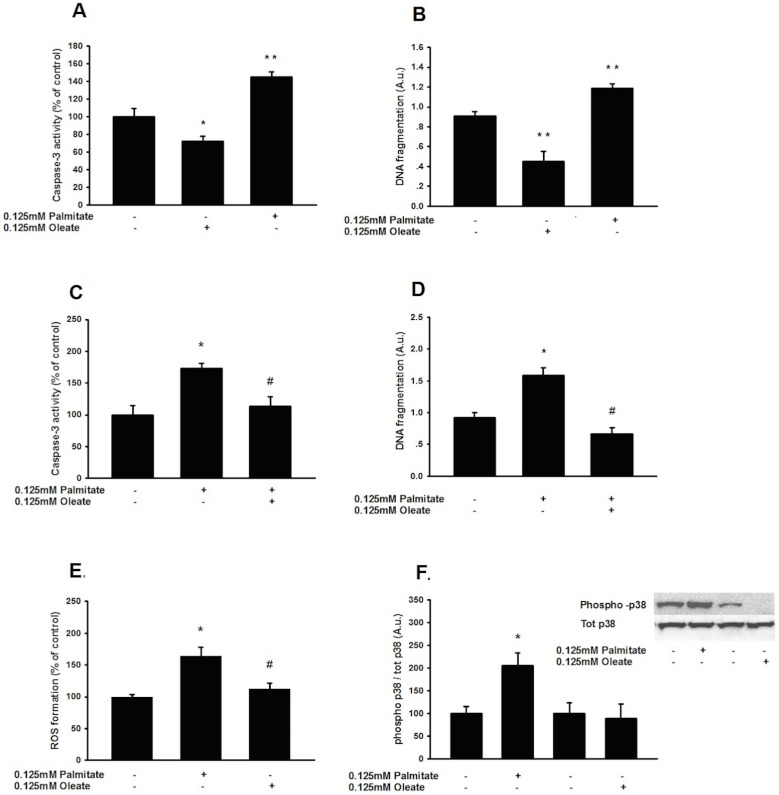
Saturated (16:0) and unsaturated (18:1) fatty acids exerted opposing effects on the induction of apoptosis (1.4-fold increase in DNA fragmentation by palmitate and a 0.5-fold reduction by oleate; p<0.01). Palmitate-induced apoptosis was associated with increased ceramide content and co-incubation with Fumonisin B1 abolished this lipo apoptosis. Oleate, on the other hand, reduced ceramide content, and-unlike palmitate-upregulated FFAR1 and FFAR3, evoking a 2-fold increase in FFAR1-mediated GLP-1 secretion following acute exposure to 0.125 mmol/L palmitate; (p<0.05).
CONCLUSION/INTERPRETATION: Saturated (16:0), but not unsaturated (18:1), fatty acids induce ceramide-mediated apoptosis of GLP-1-producing cells. Further, unsaturated fatty acids confer lipoprotection, enhancing viability and function of GLP-1-secreting cells. These data provide potential mechanistic insight contributing to reduced L-cell mass following a high fat diet and differential effects of saturated and unsaturated fatty acids on GLP-1 secretion in vivo.
脂肪酸可在体内急性刺激L细胞分泌胰高血糖素样肽-1(GLP-1)。然而,高脂饮食已被证明会降低小鼠肠道中L细胞的密度,并且L细胞数量与GLP-1分泌之间呈正相关。因此,脂肪酸刺激GLP-1分泌的机制、长期暴露于不同脂肪酸种类的升高水平的潜在影响及其潜在机制尚未完全明确。在本研究中,我们试图确定长期暴露于饱和脂肪酸(16:0)和不饱和脂肪酸(18:1)对产生GLP-1的细胞的直接影响如何改变其功能和活力以及潜在机制。
在存在/不存在饱和脂肪酸(16:0)和不饱和脂肪酸(18:1)(0.125 mM,处理48小时)的情况下培养分泌GLP-1的GLUTag细胞,随后分析细胞活力和凋亡情况,以及脂肪酸氧化、游离脂肪酸受体(FFAR1)和神经酰胺合成的参与情况。此外,还确定了对胰高血糖素原、激素原转化酶1/3(PC1/3)、游离脂肪酸受体(FFAR1、FFAR3)、钠葡萄糖共转运蛋白(SGLT)表达的影响以及随后的分泌反应。
饱和脂肪酸(16:0)和不饱和脂肪酸(18:1)对细胞凋亡的诱导产生相反的作用(棕榈酸酯使DNA片段化增加1.4倍,油酸酯使其减少0.5倍;p<0.01)。棕榈酸酯诱导的细胞凋亡与神经酰胺含量增加有关,与伏马菌素B1共同孵育可消除这种脂质凋亡。另一方面,油酸酯降低了神经酰胺含量,并且与棕榈酸酯不同,它上调了FFAR1和FFAR3,在急性暴露于0.125 mmol/L棕榈酸酯后,FFAR1介导的GLP-1分泌增加了2倍;(p<0.05)。
结论/解读:饱和脂肪酸(16:0)而非不饱和脂肪酸(18:1)可诱导产生GLP-1的细胞发生神经酰胺介导的凋亡。此外,不饱和脂肪酸具有脂质保护作用,可增强分泌GLP-1细胞的活力和功能。这些数据为高脂饮食后L细胞数量减少以及饱和脂肪酸和不饱和脂肪酸在体内对GLP-1分泌的不同影响提供了潜在的机制性见解。