Konstantinou Nikos, Pettemeridou Eva, Seimenis Ioannis, Eracleous Eleni, Papacostas Savvas S, Papanicolaou Andrew C, Constantinidou Fofi
Center for Applied Neuroscience, University of Cyprus, Nicosia, Cyprus; Department of Psychology, University of Cyprus, Nicosia, Cyprus.
Department of Medical Physics, Medical School, Democritus University of Thrace , Alexandroupolis , Greece.
Front Neurol. 2016 Mar 10;7:29. doi: 10.3389/fneur.2016.00029. eCollection 2016.
Characterize the scale and pattern of long-term atrophy in gray matter (GM), white matter (WM), and cerebrospinal fluid (CSF) in chronic moderate-severe traumatic brain injury (TBI) and its relationship to neurocognitive outcomes.
The TBI group consisted of 17 males with primary diagnosis of moderate-severe closed head injury. Participants had not received any systematic, post-acute rehabilitation and were recruited on average 8.36 years post-injury. The control group consisted of 15 males matched on age and education.
Neurocognitive battery included widely used tests of verbal memory, visual memory, executive functioning, and attention/organization. GM, WM, and CSF volumes were calculated from segmented T1-weighted anatomical MR images. Voxel-based morphometry was employed to identify brain regions with differences in GM and WM between TBI and control groups.
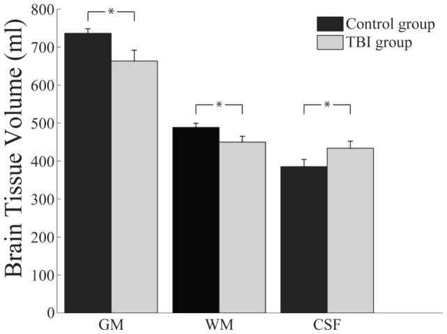
Chronic TBI results in significant neurocognitive impairments, and significant loss of GM and WM volume, and significant increase in CSF volume. Brain atrophy is not widespread, but it is rather distributed in a fronto-thalamic network. The extent of volume loss is predictive of performance on the neurocognitive tests.
Significant brain atrophy and associated neurocognitive impairments during the chronic stages of TBI support the notion that TBI results in a chronic condition with lifelong implications.
描述慢性中重度创伤性脑损伤(TBI)患者灰质(GM)、白质(WM)和脑脊液(CSF)的长期萎缩程度和模式,及其与神经认知结果的关系。
TBI组由17名男性组成,初步诊断为中重度闭合性颅脑损伤。参与者未接受过任何系统的急性后期康复治疗,平均在受伤后8.36年被招募。对照组由15名年龄和教育程度匹配的男性组成。
神经认知测试组合包括广泛使用的言语记忆、视觉记忆、执行功能以及注意力/组织能力测试。GM、WM和CSF体积通过分割后的T1加权解剖磁共振图像计算得出。基于体素的形态测量法用于识别TBI组和对照组之间GM和WM存在差异的脑区。
慢性TBI导致显著的神经认知障碍、GM和WM体积显著减少以及CSF体积显著增加。脑萎缩并不广泛,而是分布在额-丘脑网络中。体积减少的程度可预测神经认知测试的表现。
TBI慢性期显著的脑萎缩和相关神经认知障碍支持了TBI会导致一种具有终身影响的慢性疾病这一观点。