Clinical Neuropsychology Section, FGB VU University Amsterdam, Van der Boechorststraat 1, 1081BT, Amsterdam, The Netherlands.
Emma Children's Hospital, Academic Medical Center, Amsterdam, The Netherlands.
Brain Imaging Behav. 2018 Feb;12(1):29-43. doi: 10.1007/s11682-017-9673-3.
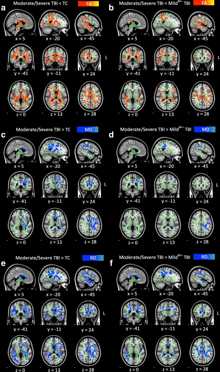
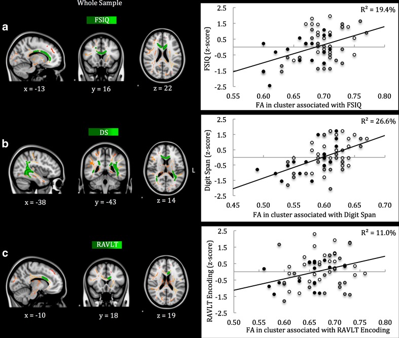
This study aims to (1) investigate the neuropathology of mild to severe pediatric TBI and (2) elucidate the predictive value of conventional and innovative neuroimaging for functional outcome. Children aged 8-14 years with trauma control (TC) injury (n = 27) were compared to children with mild TBI and risk factors for complicated TBI (mild, n = 20) or moderate/severe TBI (n = 17) at 2.8 years post-injury. Neuroimaging measures included: acute computed tomography (CT), volumetric analysis on post-acute conventional T1-weighted magnetic resonance imaging (MRI) and post-acute diffusion tensor imaging (DTI, analyzed using tract-based spatial statistics and voxel-wise regression). Functional outcome was measured using Common Data Elements for neurocognitive and behavioral functioning. The results show that intracranial pathology on acute CT-scans was more prevalent after moderate/severe TBI (65%) than after mild TBI (35%; p = .035), while both groups had decreased white matter volume on conventional MRI (ps ≤ .029, ds ≥ -0.74). The moderate/severe TBI group further showed decreased fractional anisotropy (FA) in a widespread cluster affecting all white matter tracts, in which regional associations with neurocognitive functioning were observed (FSIQ, Digit Span and RAVLT Encoding) that consistently involved the corpus callosum. FA had superior predictive value for functional outcome (i.e. intelligence, attention and working memory, encoding in verbal memory and internalizing problems) relative to acute CT-scanning (i.e. internalizing problems) and conventional MRI (no predictive value). We conclude that children with mild TBI and moderate/severe TBI are at risk of persistent white matter abnormality. Furthermore, DTI has superior predictive value for neurocognitive out-come relative to conventional neuroimaging.
(1) 研究轻度至重度儿科 TBI 的神经病理学;(2) 阐明常规和创新神经影像学对功能预后的预测价值。将创伤控制 (TC) 损伤的 8-14 岁儿童 (n=27) 与轻度 TBI 且具有复杂 TBI 危险因素的儿童 (轻度,n=20) 或中度/重度 TBI 儿童 (n=17) 进行比较。在受伤后 2.8 年进行神经影像学测量:急性计算机断层扫描 (CT),急性后常规 T1 加权磁共振成像 (MRI) 和急性后弥散张量成像 (DTI) 的容积分析(使用基于束的空间统计学和体素回归进行分析)。功能预后采用神经认知和行为功能的通用数据元素进行测量。结果表明,中度/重度 TBI 后颅内病理在急性 CT 扫描中更为常见 (65%),而轻度 TBI 后为 35%(p=0.035),但两组常规 MRI 上的白质体积均减少 (p≤0.029,ds≥-0.74)。中/重度 TBI 组进一步显示在广泛的影响所有白质束的区域内出现分数各向异性 (FA) 降低,在这些区域中观察到与神经认知功能的区域相关性 (FSIQ、数字跨度和 RAVLT 编码),这些区域均涉及胼胝体。FA 对功能预后(即智力、注意力和工作记忆、言语记忆的编码和内化问题)的预测价值优于急性 CT 扫描(即内化问题)和常规 MRI(无预测价值)。我们得出结论,轻度 TBI 和中/重度 TBI 的儿童存在持续白质异常的风险。此外,DTI 对神经认知预后的预测价值优于常规神经影像学。