Lan Martin J, Chhetry Binod Thapa, Liston Conor, Mann J John, Dubin Marc
Department of Psychiatry, Columbia University College of Physicians and Surgeons, New York, NY 10032, USA; Division of Molecular Imaging and Neuropathology, New York State Psychiatric Institute, New York, USA.
Department of Psychiatry, Columbia University College of Physicians and Surgeons, New York, NY 10032, USA; Division of Molecular Imaging and Neuropathology, New York State Psychiatric Institute, New York, USA.
Brain Stimul. 2016 Jul-Aug;9(4):577-83. doi: 10.1016/j.brs.2016.02.011. Epub 2016 Mar 2.
Repetitive transcranial magnetic stimulation (TMS) is an FDA-approved antidepressant treatment but little is known of its mechanism of action. Specifically, downstream effects of TMS remain to be elucidated.
OBJECTIVE/HYPOTHESIS: This study aims to identify brain structural changes from TMS treatment of a treatment resistant depressive episode through an exploratory analysis.
Twenty-seven subjects in a DSM-IV current major depressive episode and on a stable medication regimen had a 3T magnetic resonance T1 structural scan before and after five weeks of standard TMS treatment to the left dorsolateral prefrontal cortex. Twenty-seven healthy volunteer (HVs) subjects had the same brain MRI acquisition. Voxel-based morphometry was performed using high dimensional non-linear diffusomorphic anatomical registration (DARTEL).
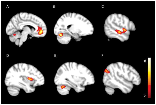
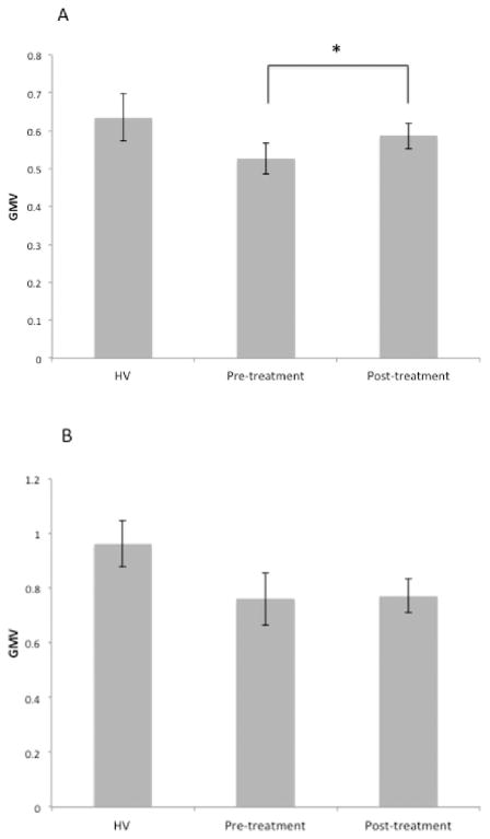
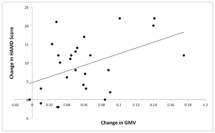
Six clusters of gray matter volume (GMV) that were lower in pre-treatment MRIs of depressed subjects than in HVs. GMV in four of these regions increased in MDD after TMS treatment by 3.5-11.2%. The four brain regions that changed with treatment were centered in the left anterior cingulate cortex, the left insula, the left superior temporal gyrus and the right angular gyrus. Increases in the anterior cingulate GMV with TMS correlated with improvement in depression severity.
To our knowledge, this is the first study of brain structural changes during TMS treatment of depression. The affected brain areas are involved in cognitive appraisal, decision-making and subjective experience of emotion. These effects may have potential relevance for the antidepressant action of TMS.
重复经颅磁刺激(TMS)是一种经美国食品药品监督管理局(FDA)批准的抗抑郁治疗方法,但其作用机制尚不清楚。具体而言,TMS的下游效应仍有待阐明。
目的/假设:本研究旨在通过探索性分析确定经TMS治疗难治性抑郁发作后的脑结构变化。
27名符合《精神疾病诊断与统计手册》第四版(DSM-IV)标准的当前重度抑郁发作且药物治疗方案稳定的受试者,在对左侧背外侧前额叶皮层进行为期五周的标准TMS治疗前后,接受了3T磁共振T1结构扫描。27名健康志愿者(HV)受试者也进行了相同的脑部MRI扫描。使用高维非线性扩散形态解剖配准(DARTEL)进行基于体素的形态测量。
抑郁受试者治疗前MRI中的灰质体积(GMV)有六个簇低于健康志愿者。经TMS治疗后,重度抑郁症(MDD)患者中这四个区域的GMV增加了3.5 - 11.2%。四个随治疗发生变化的脑区集中在左侧前扣带回皮层、左侧岛叶、左侧颞上回和右侧角回。前扣带回GMV随TMS的增加与抑郁严重程度的改善相关。
据我们所知,这是第一项关于TMS治疗抑郁症期间脑结构变化的研究。受影响的脑区涉及认知评估、决策和情绪的主观体验。这些效应可能与TMS的抗抑郁作用具有潜在相关性。