Varga Janos, Palinkas Attila, Lajko Imre, Horváth Ildikó, Boda Krisztina, Somfay Attila
Department of Pulmonology, University of Szeged, Deszk, Hungary; National Koranyi Institute for TB and Pulmonology, Budapest, Hungary.
Department of Internal Medicine, Elisabeth Hospital, Hodmezovasarhely, Hungary.
Open Respir Med J. 2016 Jan 29;10:1-11. doi: 10.2174/1874306401610010001. eCollection 2016.
The non-invasive assessment of pulmonary haemodynamics during exercise provides complementary data for the evaluation of exercise tolerance in patients with COPD.
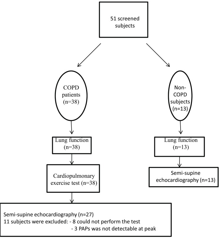
Exercise echocardiography in the semi-supine position was performed in 27 patients with COPD (C) with a forced expiratory volume in one second (FEV1) of 36±12% predicted and 13 age and gender-matched non-COPD subjects (NC). COPD patients also underwent cardiopulmonary exercise testing with gas exchange detection (CPET). Furthermore, serum high sensitive C-reactive protein (hsCRP), a marker of systemic inflammation, was also measured.
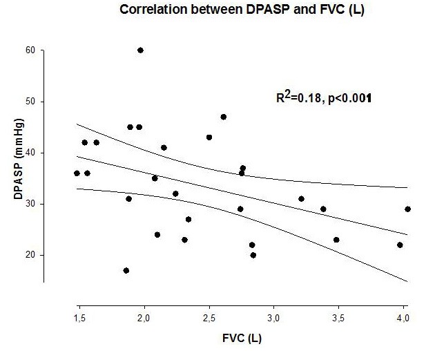
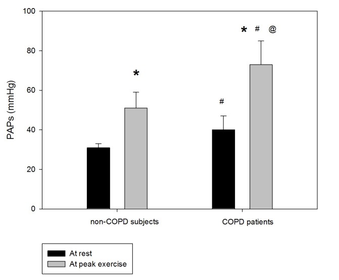
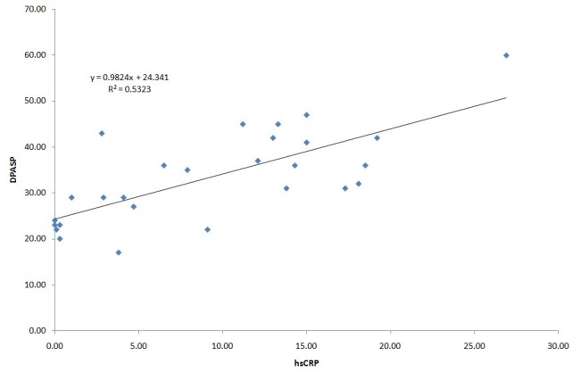
The maximal work rate (WRmax) and aerobic capacity (VO2peak) were significantly reduced (WRmax: 77±33 Watt, VO2peak: 50±14 %pred) in COPD. Pulmonary arterial systolic pressure (PAPs) was higher in COPD versus controls both at rest (39±5 vs. 31±2 mmHg, p<0.001), and at peak exercise (72±12 vs. 52±8 mmHg, p<0.001). In 19 (70%) COPD patients, the increase in PAPs was above 22 mmHg. The change in pressure (dPAPs) correlated with hsCRP (r2=0.53, p<0.0001) and forced vital capacity (FVC) (r2=0.18, p<0.001).
PAPs at rest and during exercise were significantly higher in COPD patients and correlated with higher hsCRP. This may indicate a role for systemic inflammation and hyperinflation in the pulmonary vasculature in COPD. The study was registered at ClinicalTrials.gov webpage with NCT00949195 registration number.
运动期间对肺血流动力学进行无创评估可为慢性阻塞性肺疾病(COPD)患者运动耐力的评估提供补充数据。
对27例COPD患者(C组)进行半卧位运动超声心动图检查,其一秒用力呼气容积(FEV1)为预测值的36±12%,并选取13例年龄和性别匹配的非COPD受试者(NC组)作为对照。COPD患者还接受了气体交换检测的心肺运动试验(CPET)。此外,还检测了全身炎症标志物血清高敏C反应蛋白(hsCRP)。
COPD患者的最大工作率(WRmax)和有氧能力(VO2peak)显著降低(WRmax:77±33瓦,VO2peak:50±14%预测值)。COPD患者静息时(39±5 vs. 31±2 mmHg,p<0.001)和运动峰值时(72±12 vs. 52±8 mmHg,p<0.001)的肺动脉收缩压(PAPs)均高于对照组。19例(70%)COPD患者的PAPs升高超过22 mmHg。压力变化(dPAPs)与hsCRP(r2=0.53,p<0.0001)和用力肺活量(FVC)(r2=0.18,p<0.001)相关。
COPD患者静息和运动时的PAPs显著升高,且与较高的hsCRP相关。这可能表明全身炎症和肺血管过度充气在COPD中起作用。该研究已在ClinicalTrials.gov网页注册,注册号为NCT00949195。