Lopez-Cruzan M, Sharma R, Tiwari M, Karbach S, Holstein D, Martin C R, Lechleiter J D, Herman B
Department of Cellular and Structural Biology, The University of Texas Health Science Center at San Antonio, 7703 Floyd Curl Drive, MED 238D.2, San Antonio, TX 78229-3900, USA.
Department of Cellular and Structural Biology, The University of Texas Health Science Center at San Antonio, 7703 Floyd Curl Drive, MED 238D.2, San Antonio, TX 78229-3900, USA; Center for Biomedical Neuroscience, University of Texas Health Science Center at San Antonio, San Antonio, TX 78229, USA.
Cell Death Discov. 2016 Feb 15;2:16005-. doi: 10.1038/cddiscovery.2016.5.
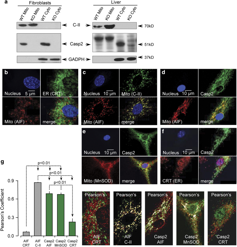
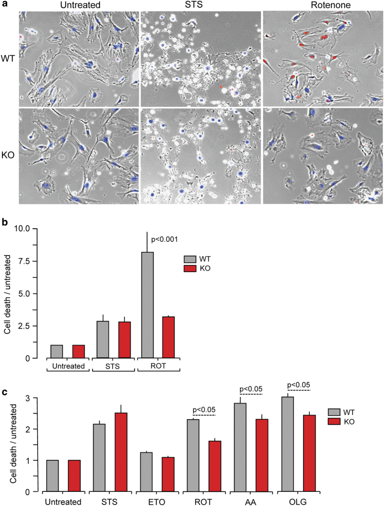
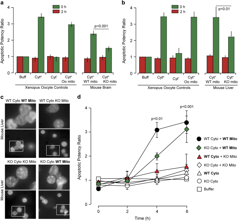
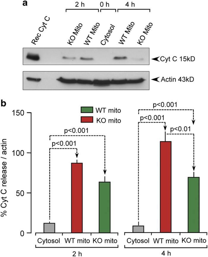
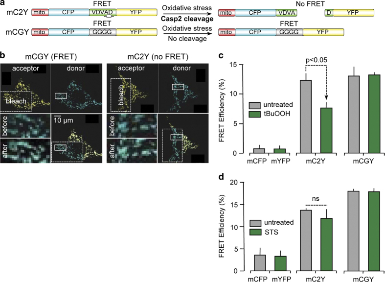
Caspase-2 plays an important role in apoptosis induced by several stimuli, including oxidative stress. However, the subcellular localization of caspase-2, particularly its presence in the mitochondria, is unclear. It is also not known if cytosolic caspase-2 translocates to the mitochondria to trigger the intrinsic pathway of apoptosis or if caspase-2 is constitutively present in the mitochondria that then selectively mediates this apoptotic effect. Here, we demonstrate the presence of caspase-2 in purified mitochondrial fractions from -cultured cells and in liver hepatocytes using immunoblots and confocal microscopy. We show that mitochondrial caspase-2 is functionally active by performing fluorescence resonance energy transfer analyses using a mitochondrially targeted substrate flanked by donor and acceptor fluorophores. Cell-free apoptotic assays involving recombination of nuclear, cytosolic and mitochondrial fractions from the livers of wild type and mice clearly point to a direct functional role for mitochondrial caspase-2 in apoptosis. Furthermore, cytochrome release from cells is decreased as compared with controls upon treatment with agents inducing mitochondrial dysfunction. Finally, we show that primary skin fibroblasts are protected from oxidants that target the mitochondrial electron transport chain. Taken together, our results demonstrate that caspase-2 exists in the mitochondria and that it is essential for mitochondrial oxidative stress-induced apoptosis.
半胱天冬酶 -2 在包括氧化应激在内的多种刺激诱导的细胞凋亡中发挥重要作用。然而,半胱天冬酶 -2 的亚细胞定位,尤其是其在线粒体中的存在情况尚不清楚。也不清楚胞质中的半胱天冬酶 -2 是否转位到线粒体以触发细胞凋亡的内在途径,或者半胱天冬酶 -2 是否组成性地存在于线粒体中,然后选择性地介导这种凋亡效应。在这里,我们使用免疫印迹和共聚焦显微镜证明了在培养细胞的纯化线粒体组分以及肝肝细胞中存在半胱天冬酶 -2。我们通过使用由供体和受体荧光团侧翼的线粒体靶向底物进行荧光共振能量转移分析,表明线粒体半胱天冬酶 -2 具有功能活性。涉及野生型和小鼠肝脏的核、胞质和线粒体组分重组的无细胞凋亡测定清楚地表明线粒体半胱天冬酶 -2 在细胞凋亡中具有直接的功能作用。此外,在用诱导线粒体功能障碍的试剂处理后,与对照相比,细胞色素从细胞中的释放减少。最后,我们表明原代皮肤成纤维细胞受到针对线粒体电子传递链的氧化剂的保护。综上所述,我们的结果表明半胱天冬酶 -2 存在于线粒体中,并且它对于线粒体氧化应激诱导的细胞凋亡至关重要。