• 文献检索
  • 文档翻译
  • 深度研究
  • 学术资讯
  • Suppr Zotero 插件Zotero 插件
  • 邀请有礼
  • 套餐&价格
  • 历史记录
应用&插件
Suppr Zotero 插件Zotero 插件浏览器插件Mac 客户端Windows 客户端微信小程序
定价
高级版会员购买积分包购买API积分包
服务
文献检索文档翻译深度研究API 文档MCP 服务
关于我们
关于 Suppr公司介绍联系我们用户协议隐私条款
关注我们

Suppr 超能文献

核心技术专利:CN118964589B侵权必究
粤ICP备2023148730 号-1Suppr @ 2026

文献检索

告别复杂PubMed语法,用中文像聊天一样搜索,搜遍4000万医学文献。AI智能推荐,让科研检索更轻松。

立即免费搜索

文件翻译

保留排版,准确专业,支持PDF/Word/PPT等文件格式,支持 12+语言互译。

免费翻译文档

深度研究

AI帮你快速写综述,25分钟生成高质量综述,智能提取关键信息,辅助科研写作。

立即免费体验

一例患有脑积水及中脑导水管进行性闭塞的人类胎儿早期综合诊断的价值:病例报告

The value of early and comprehensive diagnoses in a human fetus with hydrocephalus and progressive obliteration of the aqueduct of Sylvius: Case Report.

作者信息

Ortega Eduardo, Muñoz Rosa I, Luza Nelly, Guerra Francisco, Guerra Monserrat, Vio Karin, Henzi Roberto, Jaque Jaime, Rodriguez Sara, McAllister James P, Rodriguez Esteban

机构信息

Unidad de Neurocirugía, Instituto de Neurociencias Clínicas, Facultad de Medicina, Universidad Austral de Chile, Valdivia, Chile.

Instituto de Anatomía, Histología y Patología, Facultad de Medicina, Universidad Austral de Chile, Valdivia, Chile.

出版信息

BMC Neurol. 2016 Apr 11;16:45. doi: 10.1186/s12883-016-0566-7.

DOI:10.1186/s12883-016-0566-7
PMID:27067115
原文链接:https://pmc.ncbi.nlm.nih.gov/articles/PMC4828774/
Abstract

BACKGROUND

Mutant rodent models have highlighted the importance of the ventricular ependymal cells and the subcommissural organ (a brain gland secreting glycoproteins into the cerebrospinal fluid) in the development of fetal onset hydrocephalus. Evidence indicates that communicating and non-communicating hydrocephalus can be two sequential phases of a single pathological phenomenon triggered by ependymal disruption and/or abnormal function of the subcommissural organ. We have hypothesized that a similar phenomenon may occur in human cases with fetal onset hydrocephalus.

CASE PRESENTATION

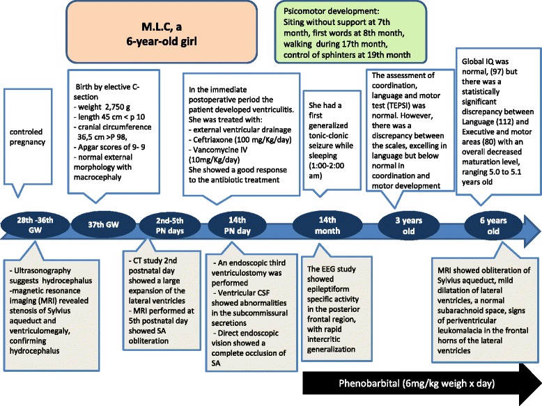
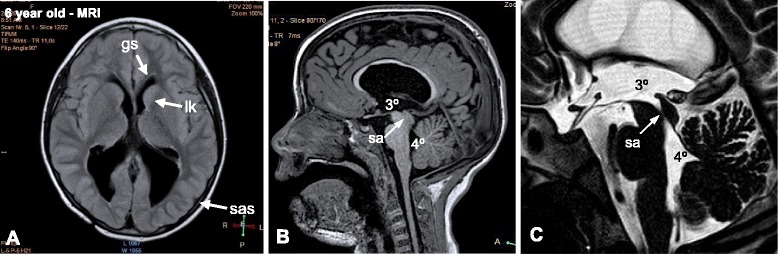
We report here on a case of human fetal communicating hydrocephalus with no central nervous system abnormalities other than stenosis of the aqueduct of Sylvius (SA) that became non-communicating hydrocephalus during the first postnatal week due to obliteration of the cerebral aqueduct. The case was followed closely by a team of basic and clinic investigators allowing an early diagnosis and prediction of the evolving pathophysiology. This information prompted neurosurgeons to perform a third ventriculostomy at postnatal day 14. The fetus was monitored by ultrasound, computerized axial tomography and magnetic resonance imaging (MRI). After birth, the follow up was by MRI, electroencephalography and neurological and neurocognitive assessments. Cerebrospinal fluid (CSF) collected at surgery showed abnormalities in the subcommissural organ proteins and the membrane proteins L1-neural cell adhesion molecule and aquaporin-4. The neurological and neurocognitive assessments at 3 and 6 years of age showed neurological impairments (epilepsy and cognitive deficits).

CONCLUSIONS

(1) In a hydrocephalic fetus, a stenosed SA can become obliterated at perinatal stages. (2) In the case reported, a close follow up of a communicating hydrocephalus detected in utero allowed a prompt postnatal surgery aiming to avoid as much brain damage as possible. (3) The clinical and pathological evolution of this patient supports the possibility that the progressive stenosis of the SA initiated during the embryonic period may have resulted from ependymal disruption of the cerebral aqueduct and dysfunction of the subcommissural organ. The analysis of subcommissural organ glycoproteins present in the CSF may be a valuable diagnostic tool for the pathogenesis of congenital hydrocephalus.

摘要

背景

突变啮齿动物模型突出了脑室室管膜细胞和联合下器官(一种向脑脊液中分泌糖蛋白的脑腺)在胎儿期脑积水发展中的重要性。有证据表明,交通性脑积水和非交通性脑积水可能是由室管膜破坏和/或联合下器官功能异常引发的单一病理现象的两个连续阶段。我们推测,类似现象可能发生在胎儿期脑积水的人类病例中。

病例报告

我们在此报告一例人类胎儿交通性脑积水病例,除中脑导水管(SA)狭窄外无其他中枢神经系统异常,该胎儿在出生后第一周因中脑导水管闭塞而变为非交通性脑积水。一个基础和临床研究团队对该病例进行了密切随访,从而实现了早期诊断并预测了不断演变的病理生理学过程。这些信息促使神经外科医生在出生后第14天进行了第三脑室造瘘术。通过超声、计算机断层扫描和磁共振成像(MRI)对胎儿进行监测。出生后,通过MRI、脑电图以及神经和神经认知评估进行随访。手术时收集的脑脊液显示联合下器官蛋白以及膜蛋白L1-神经细胞黏附分子和水通道蛋白-4存在异常。3岁和6岁时的神经和神经认知评估显示存在神经功能障碍(癫痫和认知缺陷)。

结论

(1)在脑积水胎儿中,狭窄的SA在围产期可能会闭塞。(2)在本报告的病例中,对产前检测到的交通性脑积水进行密切随访,使得能够在出生后迅速进行手术,旨在尽可能避免脑损伤。(3)该患者的临床和病理演变支持这样一种可能性,即胚胎期开始的SA进行性狭窄可能是由于中脑导水管的室管膜破坏和联合下器官功能障碍所致。对脑脊液中联合下器官糖蛋白的分析可能是先天性脑积水发病机制的一种有价值的诊断工具。

https://cdn.ncbi.nlm.nih.gov/pmc/blobs/1c11/4828774/6b4b6d6e30cd/12883_2016_566_Fig5_HTML.jpg
https://cdn.ncbi.nlm.nih.gov/pmc/blobs/1c11/4828774/94016e51d5da/12883_2016_566_Fig1_HTML.jpg
https://cdn.ncbi.nlm.nih.gov/pmc/blobs/1c11/4828774/1fc5997c0bc0/12883_2016_566_Fig2_HTML.jpg
https://cdn.ncbi.nlm.nih.gov/pmc/blobs/1c11/4828774/3b601f8b8568/12883_2016_566_Fig3_HTML.jpg
https://cdn.ncbi.nlm.nih.gov/pmc/blobs/1c11/4828774/e0f3f97ff4e1/12883_2016_566_Fig4_HTML.jpg
https://cdn.ncbi.nlm.nih.gov/pmc/blobs/1c11/4828774/6b4b6d6e30cd/12883_2016_566_Fig5_HTML.jpg

相似文献

1
The value of early and comprehensive diagnoses in a human fetus with hydrocephalus and progressive obliteration of the aqueduct of Sylvius: Case Report.一例患有脑积水及中脑导水管进行性闭塞的人类胎儿早期综合诊断的价值:病例报告
BMC Neurol. 2016 Apr 11;16:45. doi: 10.1186/s12883-016-0566-7.
2
Cellular mechanisms involved in the stenosis and obliteration of the cerebral aqueduct of hyh mutant mice developing congenital hydrocephalus.参与先天性脑积水的hyh突变小鼠大脑导水管狭窄和闭塞的细胞机制。
J Neuropathol Exp Neurol. 2003 Oct;62(10):1019-40. doi: 10.1093/jnen/62.10.1019.
3
Role of the subcommissural organ in the pathogenesis of congenital hydrocephalus in the HTx rat.Subcommissural Organ 在 HTx 大鼠先天性脑积水发病机制中的作用。
Cell Tissue Res. 2013 Jun;352(3):707-25. doi: 10.1007/s00441-013-1615-9. Epub 2013 May 4.
4
Spontaneous congenital hydrocephalus in the mutant mouse hyh. Changes in the ventricular system and the subcommissural organ.突变小鼠hyh中的自发性先天性脑积水。脑室系统和联合下器官的变化。
J Neuropathol Exp Neurol. 1998 Feb;57(2):188-202. doi: 10.1097/00005072-199802000-00010.
5
Hydrocephalus induced by immunological blockage of the subcommissural organ-Reissner's fiber (RF) complex by maternal transfer of anti-RF antibodies.通过母体转移抗Reissner纤维(RF)抗体对联合下器官 - Reissner纤维(RF)复合体进行免疫阻断所诱导的脑积水。
Exp Brain Res. 2000 Nov;135(1):41-52. doi: 10.1007/s002210000474.
6
Reduced subcommissural organ glycoprotein immunoreactivity precedes aqueduct closure and ventricular dilatation in H-Tx rat hydrocephalus.在H-Tx大鼠脑积水模型中,连合下器官糖蛋白免疫反应性降低先于导水管闭合和脑室扩张。
Cell Tissue Res. 2004 Mar;315(3):361-73. doi: 10.1007/s00441-003-0843-9. Epub 2004 Jan 14.
7
A programmed ependymal denudation precedes congenital hydrocephalus in the hyh mutant mouse.在hyh突变小鼠中,程序性室管膜剥脱先于先天性脑积水出现。
J Neuropathol Exp Neurol. 2001 Nov;60(11):1105-19. doi: 10.1093/jnen/60.11.1105.
8
Bi-allelic variations in CRB2, encoding the crumbs cell polarity complex component 2, lead to non-communicating hydrocephalus due to atresia of the aqueduct of sylvius and central canal of the medulla.CRB2 基因的双等位基因突变导致非交通性脑积水,其病因是西尔维乌斯导水管和延髓中央管狭窄。CRB2 基因编码crumbs 细胞极性复合物的组成部分 2。
Acta Neuropathol Commun. 2023 Feb 20;11(1):29. doi: 10.1186/s40478-023-01519-8.
9
Subcommissural organ dysfunction in H-Tx rats with early-onset hydrocephalus.早发性脑积水H-Tx大鼠的联合下器官功能障碍
Eur J Pediatr Surg. 2002 Dec;12 Suppl 1:S45-7.
10
Congenital hydrocephalus following X-irradiation of pregnant rats on an early gestational day.妊娠早期接受X射线照射的孕鼠所患先天性脑积水。
Neurobehav Toxicol Teratol. 1986 Mar-Apr;8(2):143-50.

引用本文的文献

1
C-mannosyltransferase DPY19L1L-mediated Reissner Fiber formation is critical for zebrafish () body axis straightening.C-甘露糖基转移酶DPY19L1L介导的Reissner纤维形成对斑马鱼体轴伸直至关重要。
Sci Adv. 2025 May 9;11(19):eadv2032. doi: 10.1126/sciadv.adv2032.
2
Congenital hydrocephalus: a review of recent advances in genetic etiology and molecular mechanisms.先天性脑积水:遗传病因学和分子机制研究进展综述。
Mil Med Res. 2024 Aug 12;11(1):54. doi: 10.1186/s40779-024-00560-5.
3
The subcommissural organ regulates brain development via secreted peptides.

本文引用的文献

1
Cell Junction Pathology of Neural Stem Cells Is Associated With Ventricular Zone Disruption, Hydrocephalus, and Abnormal Neurogenesis.神经干细胞的细胞连接病理学与脑室区破坏、脑积水及异常神经发生相关。
J Neuropathol Exp Neurol. 2015 Jul;74(7):653-71. doi: 10.1097/NEN.0000000000000203.
2
Genetic deletion of afadin causes hydrocephalus by destruction of adherens junctions in radial glial and ependymal cells in the midbrain.afadin 基因缺失通过破坏中脑放射状胶质细胞和室管膜细胞中的黏着连接导致脑积水。
PLoS One. 2013 Nov 13;8(11):e80356. doi: 10.1371/journal.pone.0080356. eCollection 2013.
3
Disruption of CDH2/N-cadherin-based adherens junctions leads to apoptosis of ependymal cells and denudation of brain ventricular walls.
室下器官通过分泌的肽调节大脑发育。
Nat Neurosci. 2024 Jun;27(6):1103-1115. doi: 10.1038/s41593-024-01639-x. Epub 2024 May 13.
4
The subcommissural organ regulates brain development via secreted peptides.连合下器官通过分泌肽来调节大脑发育。
bioRxiv. 2024 Mar 31:2024.03.30.587415. doi: 10.1101/2024.03.30.587415.
5
SCO-spondin knockout mice exhibit small brain ventricles and mild spine deformation.SCO-spondin 敲除小鼠表现出脑室内小和轻微的脊柱变形。
Fluids Barriers CNS. 2023 Dec 5;20(1):89. doi: 10.1186/s12987-023-00491-8.
6
Severe Phenotype in Patients with X-linked Hydrocephalus Caused by a Missense Mutation in L1CAM.L1CAM基因错义突变导致的X连锁脑积水患者的严重表型
Turk Arch Pediatr. 2022 Sep;57(5):521-525. doi: 10.5152/TurkArchPediatr.2022.22070.
7
Turning the Curve Into Straight: Phenogenetics of the Spine Morphology and Coordinate Maintenance in the Zebrafish.化曲为直:斑马鱼脊柱形态与坐标维持的表型遗传学
Front Cell Dev Biol. 2022 Jan 26;9:801652. doi: 10.3389/fcell.2021.801652. eCollection 2021.
8
SCO-spondin, a giant matricellular protein that regulates cerebrospinal fluid activity.SCO-spondin,一种调节脑脊液活动的巨大细胞外基质蛋白。
Fluids Barriers CNS. 2021 Oct 2;18(1):45. doi: 10.1186/s12987-021-00277-w.
9
Immune activation during brain infection in African infants with frequent cytomegalovirus co-infection.在频繁合并巨细胞病毒感染的非洲婴儿脑部感染期间的免疫激活。
iScience. 2021 Mar 23;24(4):102351. doi: 10.1016/j.isci.2021.102351. eCollection 2021 Apr 23.
10
Role of aquaporins in hydrocephalus: what do we know and where do we stand? A systematic review.水通道蛋白在脑积水中的作用:我们了解多少,现状如何?系统综述。
J Neurol. 2021 Nov;268(11):4078-4094. doi: 10.1007/s00415-020-10122-z. Epub 2020 Aug 3.
CDH2/N-钙黏蛋白黏着连接的破坏导致室管膜细胞凋亡和脑室内壁剥脱。
J Neuropathol Exp Neurol. 2013 Sep;72(9):846-60. doi: 10.1097/NEN.0b013e3182a2d5fe.
4
Aquaporin-4 expression in the cerebrospinal fluid in congenital human hydrocephalus.水通道蛋白-4 在先天性人类脑积水脑脊液中的表达。
Fluids Barriers CNS. 2013 May 9;10(1):18. doi: 10.1186/2045-8118-10-18.
5
Role of the subcommissural organ in the pathogenesis of congenital hydrocephalus in the HTx rat.Subcommissural Organ 在 HTx 大鼠先天性脑积水发病机制中的作用。
Cell Tissue Res. 2013 Jun;352(3):707-25. doi: 10.1007/s00441-013-1615-9. Epub 2013 May 4.
6
A cell junction pathology of neural stem cells leads to abnormal neurogenesis and hydrocephalus.神经干细胞的细胞连接病理学导致异常神经发生和脑积水。
Biol Res. 2012;45(3):231-42. doi: 10.4067/S0716-97602012000300005.
7
Pediatric hydrocephalus outcomes: a review.小儿脑积水的结局:综述。
Fluids Barriers CNS. 2012 Aug 27;9(1):18. doi: 10.1186/2045-8118-9-18.
8
Alterations in protein regulators of neurodevelopment in the cerebrospinal fluid of infants with posthemorrhagic hydrocephalus of prematurity.早产儿出血后脑积水患儿脑脊液中神经发育蛋白调节因子的改变。
Mol Cell Proteomics. 2012 Jun;11(6):M111.011973. doi: 10.1074/mcp.M111.011973. Epub 2011 Dec 20.
9
Neuroependymal denudation is in progress in full-term human foetal spina bifida aperta.足月开放性脊柱裂的人类胎儿正在发生神经室管膜剥脱。
Brain Pathol. 2011 Mar;21(2):163-79. doi: 10.1111/j.1750-3639.2010.00432.x.
10
Malformations of cortical development and aberrant cortical networks: epileptogenesis and functional organization.皮质发育畸形和皮质网络异常:致痫性和功能组织。
J Clin Neurophysiol. 2010 Dec;27(6):372-9. doi: 10.1097/WNP.0b013e3181fe0585.