van der Hoeven Barbara, Oudshoorn Diede, Koster Abraham J, Snijder Eric J, Kikkert Marjolein, Bárcena Montserrat
Electron Microscopy Section, Department of Molecular Cell Biology, Leiden University Medical Center, Leiden, The Netherlands.
Molecular Virology Laboratory, Department of Medical Microbiology, Leiden University Medical Center, Leiden, The Netherlands.
Virus Res. 2016 Jul 15;220:70-90. doi: 10.1016/j.virusres.2016.04.001. Epub 2016 Apr 9.
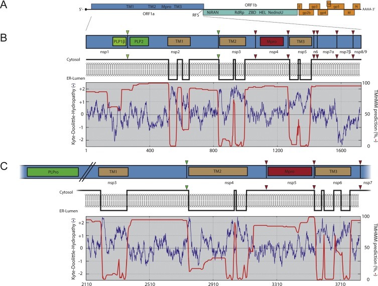
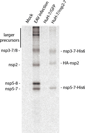
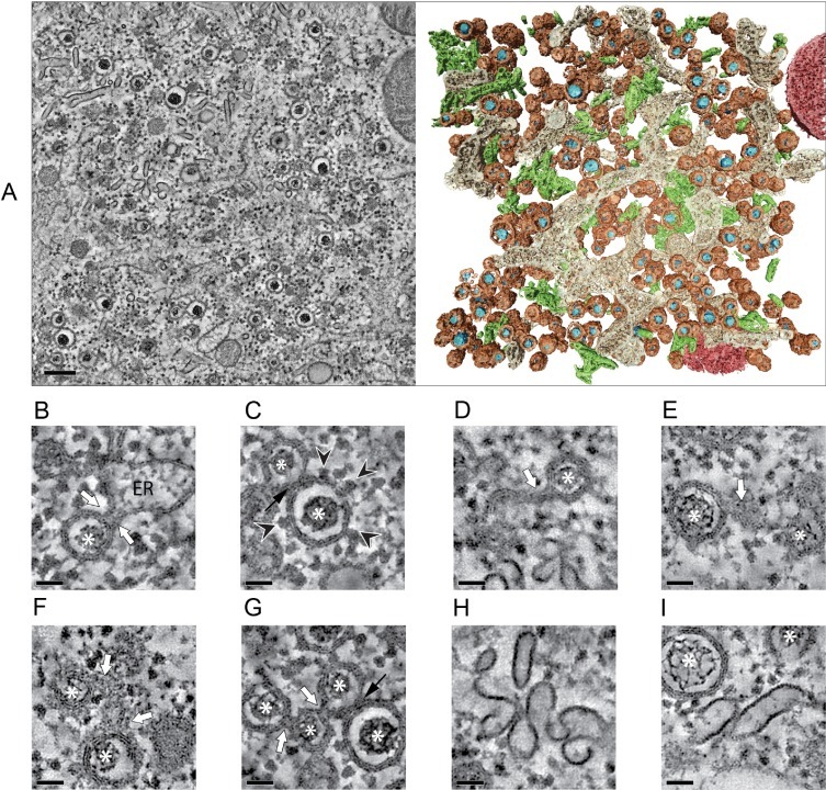
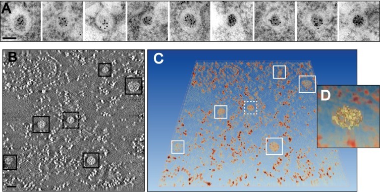
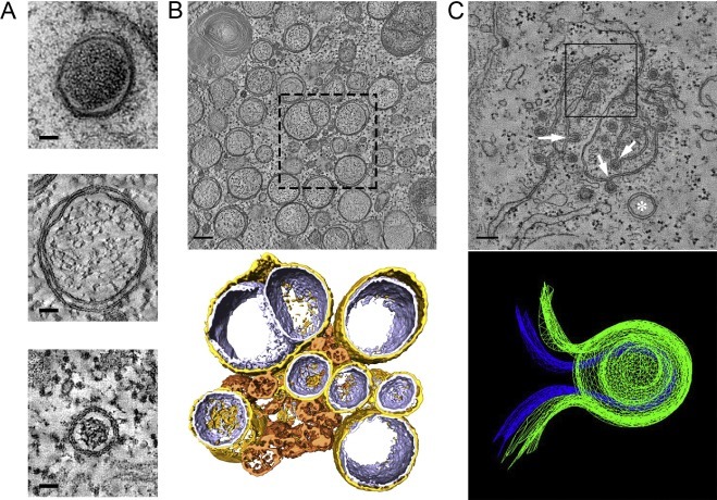
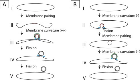
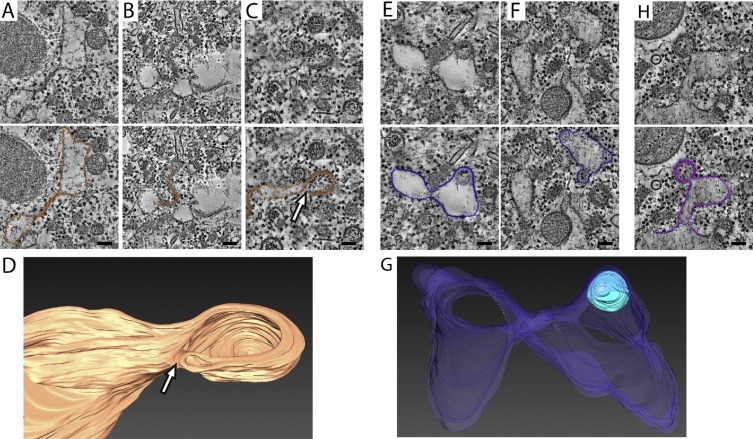
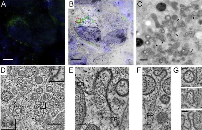
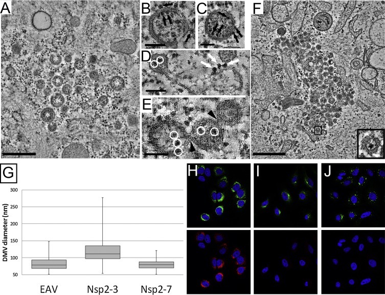
All eukaryotic positive-stranded RNA (+RNA) viruses appropriate host cell membranes and transform them into replication organelles, specialized micro-environments that are thought to support viral RNA synthesis. Arteriviruses (order Nidovirales) belong to the subset of +RNA viruses that induce double-membrane vesicles (DMVs), similar to the structures induced by e.g. coronaviruses, picornaviruses and hepatitis C virus. In the last years, electron tomography has revealed substantial differences between the structures induced by these different virus groups. Arterivirus-induced DMVs appear to be closed compartments that are continuous with endoplasmic reticulum membranes, thus forming an extensive reticulovesicular network (RVN) of intriguing complexity. This RVN is remarkably similar to that described for the distantly related coronaviruses (also order Nidovirales) and sets them apart from other DMV-inducing viruses analysed to date. We review here the current knowledge and open questions on arterivirus replication organelles and discuss them in the light of the latest studies on other DMV-inducing viruses, particularly coronaviruses. Using the equine arteritis virus (EAV) model system and electron tomography, we present new data regarding the biogenesis of arterivirus-induced DMVs and uncover numerous putative intermediates in DMV formation. We generated cell lines that can be induced to express specific EAV replicase proteins and showed that DMVs induced by the transmembrane proteins nsp2 and nsp3 form an RVN and are comparable in topology and architecture to those formed during viral infection. Co-expression of the third EAV transmembrane protein (nsp5), expressed as part of a self-cleaving polypeptide that mimics viral polyprotein processing in infected cells, led to the formation of DMVs whose size was more homogenous and closer to what is observed upon EAV infection, suggesting a regulatory role for nsp5 in modulating membrane curvature and DMV formation.
所有真核生物正链RNA(+RNA)病毒都会占据合适的宿主细胞膜,并将其转化为复制细胞器,即被认为可支持病毒RNA合成的特殊微环境。动脉炎病毒(尼多病毒目)属于+RNA病毒的一个亚群,可诱导形成双膜囊泡(DMV),这与例如冠状病毒、小RNA病毒和丙型肝炎病毒所诱导形成的结构相似。在过去几年中,电子断层扫描揭示了这些不同病毒组所诱导形成的结构之间存在显著差异。动脉炎病毒诱导形成的DMV似乎是与内质网膜连续的封闭隔室,从而形成了一个复杂程度令人着迷的广泛网状囊泡网络(RVN)。这个RVN与远亲冠状病毒(也属于尼多病毒目)所描述的网络非常相似,并且使其有别于迄今为止分析过的其他诱导DMV形成的病毒。我们在此回顾关于动脉炎病毒复制细胞器的现有知识和未解决的问题,并根据对其他诱导DMV形成的病毒,特别是冠状病毒的最新研究进行讨论。利用马动脉炎病毒(EAV)模型系统和电子断层扫描,我们给出了关于动脉炎病毒诱导形成的DMV生物发生的新数据,并发现了DMV形成过程中的众多假定中间体。我们构建了可被诱导表达特定EAV复制酶蛋白的细胞系,并表明由跨膜蛋白nsp2和nsp3诱导形成的DMV形成了一个RVN,其拓扑结构和结构与病毒感染期间形成的DMV相当。第三个EAV跨膜蛋白(nsp5)作为一种自切割多肽的一部分表达,该多肽模拟感染细胞中的病毒多蛋白加工过程,其共表达导致形成大小更均匀且更接近EAV感染时所观察到的DMV,这表明nsp5在调节膜曲率和DMV形成中具有调节作用。