• 文献检索
  • 文档翻译
  • 深度研究
  • 学术资讯
  • Suppr Zotero 插件Zotero 插件
  • 邀请有礼
  • 套餐&价格
  • 历史记录
应用&插件
Suppr Zotero 插件Zotero 插件浏览器插件Mac 客户端Windows 客户端微信小程序
定价
高级版会员购买积分包购买API积分包
服务
文献检索文档翻译深度研究API 文档MCP 服务
关于我们
关于 Suppr公司介绍联系我们用户协议隐私条款
关注我们

Suppr 超能文献

核心技术专利:CN118964589B侵权必究
粤ICP备2023148730 号-1Suppr @ 2026

文献检索

告别复杂PubMed语法,用中文像聊天一样搜索,搜遍4000万医学文献。AI智能推荐,让科研检索更轻松。

立即免费搜索

文件翻译

保留排版,准确专业,支持PDF/Word/PPT等文件格式,支持 12+语言互译。

免费翻译文档

深度研究

AI帮你快速写综述,25分钟生成高质量综述,智能提取关键信息,辅助科研写作。

立即免费体验

视神经鞘作为眼球运动中眼球上一种新的机械负荷。

Optic Nerve Sheath as a Novel Mechanical Load on the Globe in Ocular Duction.

作者信息

Demer Joseph L

出版信息

Invest Ophthalmol Vis Sci. 2016 Apr;57(4):1826-38. doi: 10.1167/iovs.15-18718.

DOI:10.1167/iovs.15-18718
PMID:27082297
原文链接:https://pmc.ncbi.nlm.nih.gov/articles/PMC4849549/
Abstract

PURPOSE

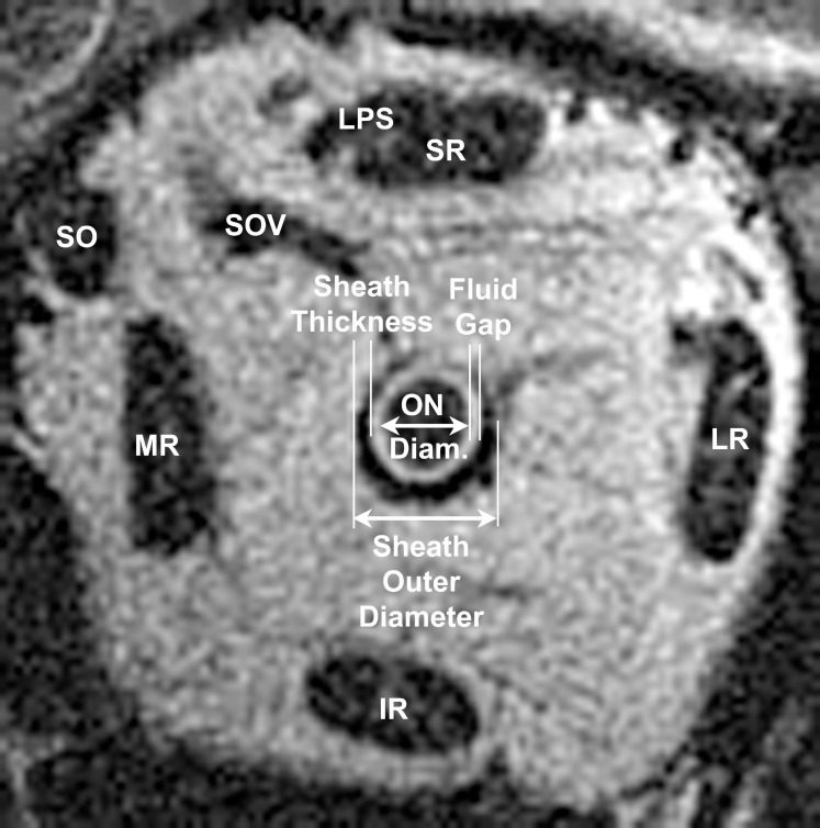
The optic nerve (ON) sheath's role in limiting duction has been previously unappreciated. This study employed magnetic resonance imaging (MRI) to demonstrate this constraint on adduction.

METHODS

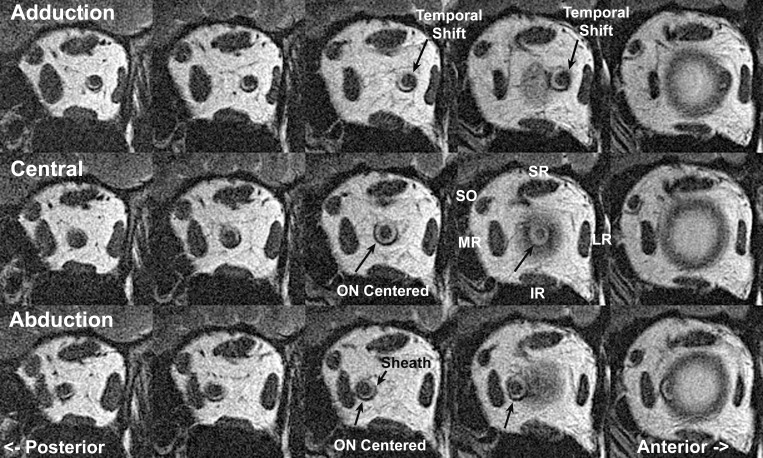
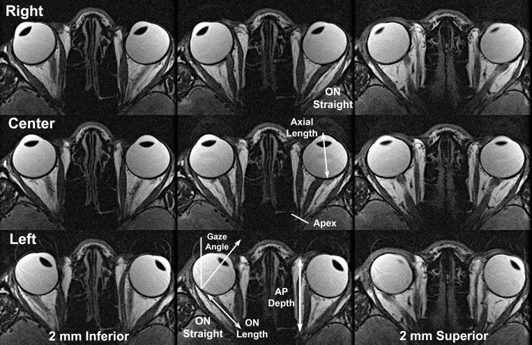
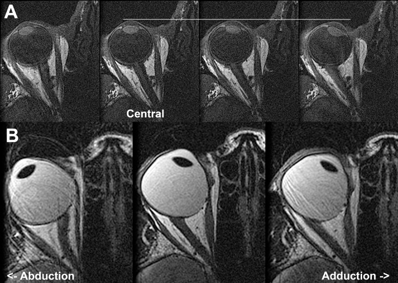
High-resolution, surface coil axial MRI was obtained in 11 normal adults, 14 subjects with esotropia (ET) having normal axial length (AL) < 25.8 mm, 13 myopic subjects with ET and mean AL 29.3 ± 3.3 (SD) mm, and 7 subjects with exotropia (XT). Gaze angles and ON lengths were measured for scans employing eccentric lateral fixation in which an ON became completely straightened.

RESULTS

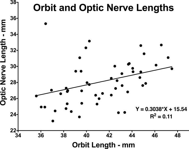
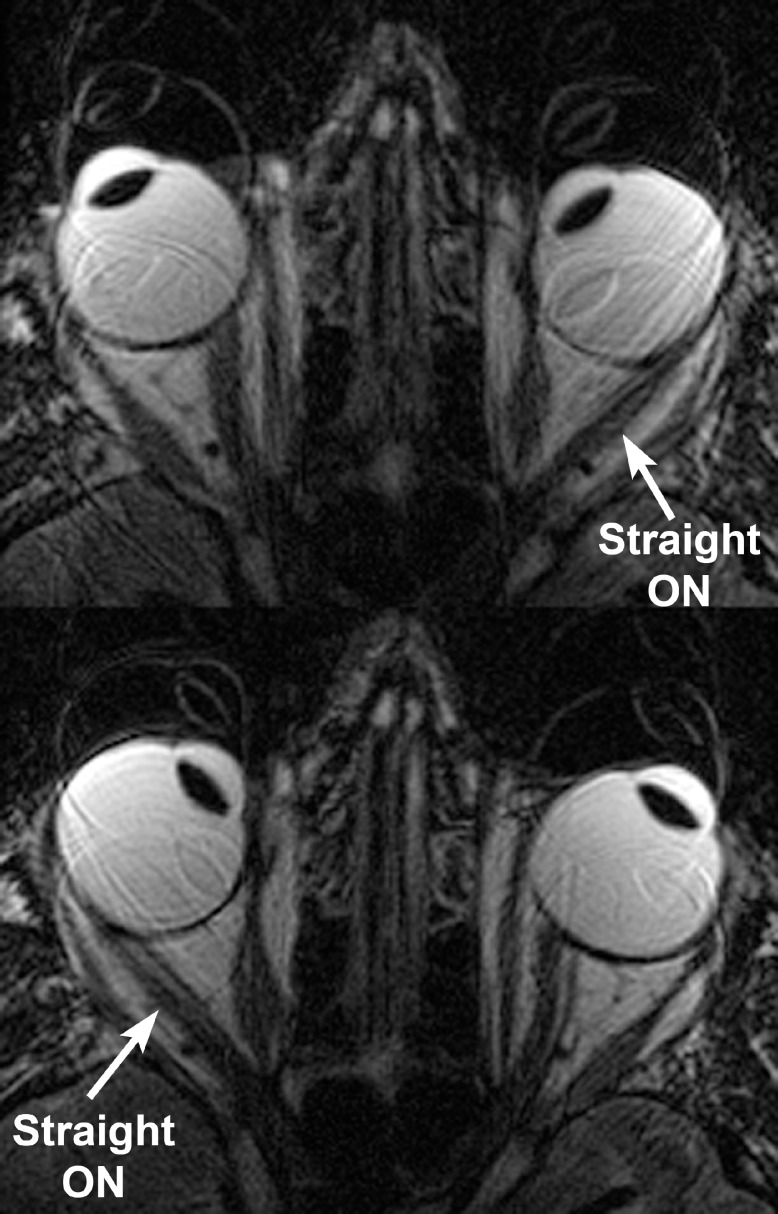
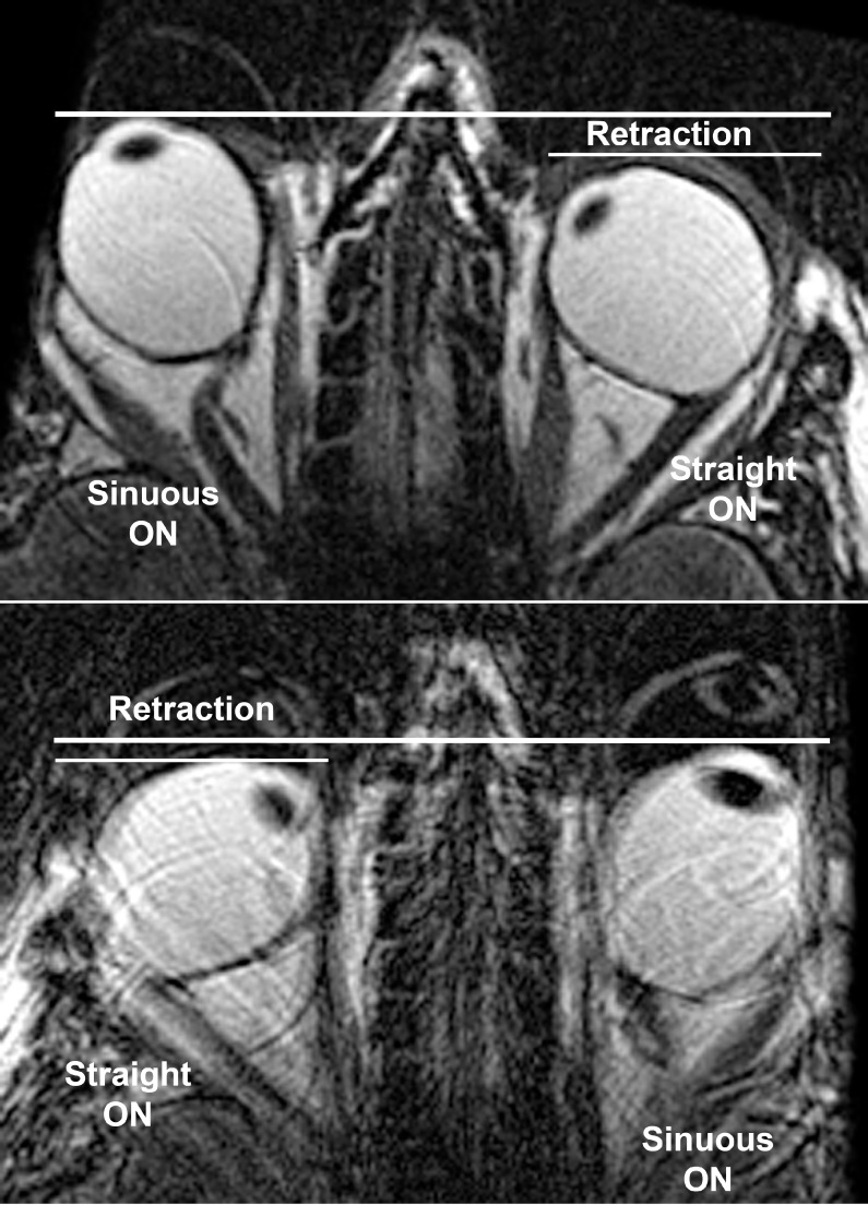
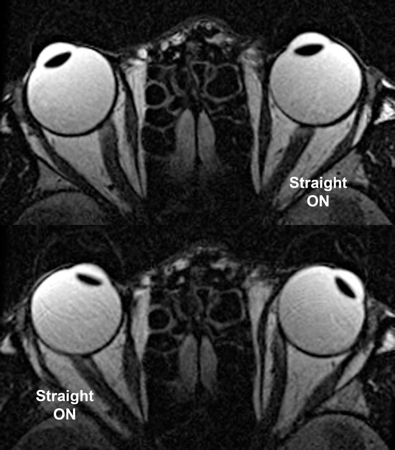
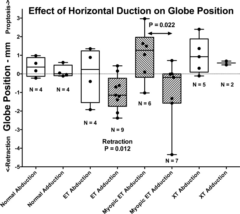
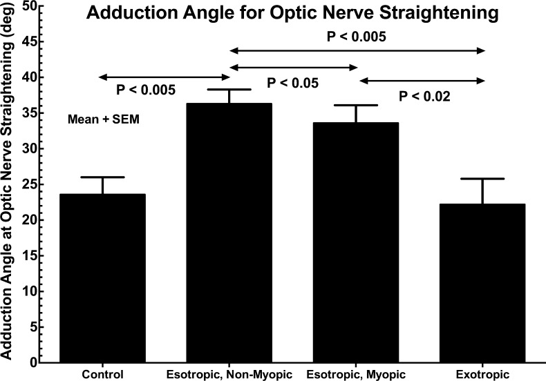
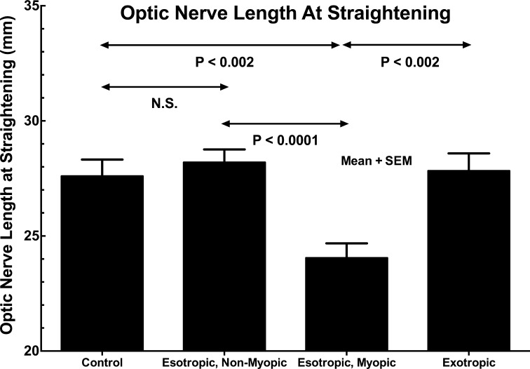
In all groups, ON straightening occurred only in the adducting, not abducting, eye. Adduction at ON straightening was 26.0 ± 8.8° in normal subjects, not significantly different from XT at 22.2 ± 11.8°. However, there was significant increase in comparable adduction in ET to 36.3 ± 9.3°, and in myopic ET to 33.6 ± 10.7° (P < 0.04). Optic nerve length at straightening was 27.6 ± 2.7 mm in normals, not significantly different from 28.2 ± 2.8 mm in ET and 27.8 ± 2.7 mm in XT. In myopic ET, ON length at straightening was significantly reduced to 24.0 ± 2.9 mm (P < 0.002) and was associated with globe retraction in adduction, suggesting ON tethering.

CONCLUSIONS

Large adduction may exhaust length redundancy in the normally sinuous ON and sheath, so that additional adduction must stretch the sheath and retract or deform the globe. These mechanical effects are most significant in ET with axial myopia, but may also exert traction on the posterior sclera absent strabismus or myopia. Tethering by the ON sheath in adduction is an important, novel mechanical load on the globe.

摘要

目的

视神经鞘在限制眼球转动方面的作用此前未得到充分认识。本研究采用磁共振成像(MRI)来证明其对内收的这种限制。

方法

对11名正常成年人、14名轴向长度(AL)正常(<25.8 mm)的内斜视(ET)患者、13名患有ET且平均AL为29.3±3.3(标准差)mm的近视患者以及7名外斜视(XT)患者进行了高分辨率表面线圈轴向MRI检查。对于采用偏心外侧注视使视神经完全伸直的扫描,测量注视角度和视神经长度。

结果

在所有组中,视神经伸直仅发生在内收眼,而非外展眼。正常受试者视神经伸直时的内收角度为26.0±8.8°,与XT的22.2±11.8°无显著差异。然而,ET的可比内收角度显著增加至36.3±9.3°,近视性ET则增加至33.6±10.7°(P<0.04)。正常情况下视神经伸直时的长度为27.6±2.7 mm,与ET的28.2±2.8 mm和XT的27.8±2.7 mm无显著差异。在近视性ET中,视神经伸直时的长度显著缩短至24.0±2.9 mm(P<0.002),且与内收时眼球后缩相关,提示视神经受束缚。

结论

大幅度内收可能耗尽正常呈弯曲状的视神经及其鞘的长度冗余,因此额外的内收必须拉伸鞘并使眼球后缩或变形。这些机械效应在伴有轴性近视的ET中最为显著,但在无斜视或近视的情况下也可能对后巩膜施加牵引力。内收时视神经鞘的束缚是眼球上一种重要的新的机械负荷。

https://cdn.ncbi.nlm.nih.gov/pmc/blobs/d1be/4849549/158349b23e9c/i1552-5783-57-4-1826-f11.jpg
https://cdn.ncbi.nlm.nih.gov/pmc/blobs/d1be/4849549/32c08075e48b/i1552-5783-57-4-1826-f01.jpg
https://cdn.ncbi.nlm.nih.gov/pmc/blobs/d1be/4849549/1e8d77f260b6/i1552-5783-57-4-1826-f02.jpg
https://cdn.ncbi.nlm.nih.gov/pmc/blobs/d1be/4849549/57cf0b7e9e2b/i1552-5783-57-4-1826-f03.jpg
https://cdn.ncbi.nlm.nih.gov/pmc/blobs/d1be/4849549/c3b91eb29d9f/i1552-5783-57-4-1826-f04.jpg
https://cdn.ncbi.nlm.nih.gov/pmc/blobs/d1be/4849549/a3d4a5db6096/i1552-5783-57-4-1826-f05.jpg
https://cdn.ncbi.nlm.nih.gov/pmc/blobs/d1be/4849549/ebd37c9afac1/i1552-5783-57-4-1826-f06.jpg
https://cdn.ncbi.nlm.nih.gov/pmc/blobs/d1be/4849549/4d4b81357943/i1552-5783-57-4-1826-f07.jpg
https://cdn.ncbi.nlm.nih.gov/pmc/blobs/d1be/4849549/ac9e83bf5e92/i1552-5783-57-4-1826-f08.jpg
https://cdn.ncbi.nlm.nih.gov/pmc/blobs/d1be/4849549/de3ef3f299a8/i1552-5783-57-4-1826-f09.jpg
https://cdn.ncbi.nlm.nih.gov/pmc/blobs/d1be/4849549/15f5604400c6/i1552-5783-57-4-1826-f10.jpg
https://cdn.ncbi.nlm.nih.gov/pmc/blobs/d1be/4849549/158349b23e9c/i1552-5783-57-4-1826-f11.jpg

相似文献

1
Optic Nerve Sheath as a Novel Mechanical Load on the Globe in Ocular Duction.视神经鞘作为眼球运动中眼球上一种新的机械负荷。
Invest Ophthalmol Vis Sci. 2016 Apr;57(4):1826-38. doi: 10.1167/iovs.15-18718.
2
Optic Nerve Sheath Tethering in Adduction Occurs in Esotropia and Hypertropia, But Not in Exotropia.内收时视神经鞘牵拉仅见于内斜视和上斜视,而外斜视则不然。
Invest Ophthalmol Vis Sci. 2018 Jun 1;59(7):2899-2904. doi: 10.1167/iovs.18-24305.
3
Magnetic Resonance Imaging of Optic Nerve Traction During Adduction in Primary Open-Angle Glaucoma With Normal Intraocular Pressure.正常眼压原发性开角型青光眼内收时视神经牵拉的磁共振成像
Invest Ophthalmol Vis Sci. 2017 Aug 1;58(10):4114-4125. doi: 10.1167/iovs.17-22093.
4
Adduction-Induced Strain on the Optic Nerve in Primary Open Angle Glaucoma at Normal Intraocular Pressure.正常眼压原发性开角型青光眼视神经营养不良时的内收诱导性视神经应变。
Curr Eye Res. 2021 Apr;46(4):568-578. doi: 10.1080/02713683.2020.1817491. Epub 2020 Sep 11.
5
[Displacement of the posterior part of the eyeball in myopia].[近视时眼球后部的移位]
Nippon Ganka Gakkai Zasshi. 2006 Dec;110(12):984-9.
6
Heavy eye syndrome versus sagging eye syndrome in high myopia.高度近视中的重度眼综合征与下垂眼综合征
J AAPOS. 2015 Dec;19(6):500-6. doi: 10.1016/j.jaapos.2015.08.012.
7
"Heavy Eye" syndrome in the absence of high myopia: A connective tissue degeneration in elderly strabismic patients.无高度近视情况下的“沉重眼”综合征:老年斜视患者的一种结缔组织退变
J AAPOS. 2009 Feb;13(1):36-44. doi: 10.1016/j.jaapos.2008.07.008. Epub 2008 Oct 18.
8
Knobby Eye Syndrome.结节状眼综合征
Strabismus. 2018 Mar;26(1):33-41. doi: 10.1080/09273972.2017.1418393. Epub 2017 Dec 26.
9
Measurement of recti eye muscle paths by magnetic resonance imaging in highly myopic and normal subjects.通过磁共振成像测量高度近视和正常受试者的直肌眼肌路径。
Invest Ophthalmol Vis Sci. 1999 Oct;40(11):2554-60.
10
[The posterior portion of the eyeball and the muscle cone in cases of high myopia].[高度近视病例中眼球后部及肌锥]
Nippon Ganka Gakkai Zasshi. 2004 Jan;108(1):12-7.

引用本文的文献

1
Interpupillary Distance and Peripapillary Myopic Changes: A Pilot Study in a Glaucomatous Cohort.瞳距与视乳头周围近视性改变:青光眼队列的一项初步研究
J Clin Med. 2025 Jul 10;14(14):4895. doi: 10.3390/jcm14144895.
2
Impact of Optic Nerve Tortuosity, Globe Proptosis, and Size on Retinal Ganglion Cell Thickness Across General, Glaucoma, and Myopic Populations.视神经弯曲度、眼球突出度和大小对普通人群、青光眼患者和近视人群视网膜神经节细胞厚度的影响。
Invest Ophthalmol Vis Sci. 2025 Jun 2;66(6):4. doi: 10.1167/iovs.66.6.4.
3
Asymmetric stress distribution on lamina cribrosa in glaucoma patients with high myopia.

本文引用的文献

1
Optic nerve head biomechanics in aging and disease.衰老与疾病中的视神经乳头生物力学
Exp Eye Res. 2015 Apr;133:19-29. doi: 10.1016/j.exer.2015.02.011.
2
Collagen microstructural factors influencing optic nerve head biomechanics.影响视神经乳头生物力学的胶原微观结构因素
Invest Ophthalmol Vis Sci. 2015 Mar 3;56(3):2031-42. doi: 10.1167/iovs.14-15734.
3
Peripapillary choroidal thickness in young Asians with high myopia.高度近视亚洲年轻人的视乳头周围脉络膜厚度
高度近视青光眼患者筛板上的不对称应力分布。
Graefes Arch Clin Exp Ophthalmol. 2025 Mar 26. doi: 10.1007/s00417-025-06809-6.
4
Horizontal Gaze Tolerance and Its Effects on Visual Sensitivity in Glaucoma.青光眼患者的水平注视耐受及其对视觉敏感度的影响
Invest Ophthalmol Vis Sci. 2025 Jan 2;66(1):59. doi: 10.1167/iovs.66.1.59.
5
Eye Movements and the Intraorbital Subarachnoid Space: Potential Contribution of Altered Cerebrospinal Fluid Pumping in Optic Neuropathies.眼球运动与眶内蛛网膜下腔:脑脊液泵功能改变在视神经病变中的潜在作用
Invest Ophthalmol Vis Sci. 2025 Jan 2;66(1):53. doi: 10.1167/iovs.66.1.53.
6
Exploration of Choroidal Thinning Located Temporal to the Fovea: A Pilot Study.黄斑颞侧脉络膜变薄的探索:一项初步研究。
J Clin Med. 2024 Aug 23;13(17):4978. doi: 10.3390/jcm13174978.
7
Tensile properties of glaucomatous human sclera, optic nerve, and optic nerve sheath.青光眼患者巩膜、视神经和视神经鞘的拉伸性能。
Biomech Model Mechanobiol. 2024 Dec;23(6):1851-1862. doi: 10.1007/s10237-024-01872-0. Epub 2024 Aug 8.
8
Postmortem Digital Image Correlation and Finite Element Modeling Demonstrate Posterior Scleral Deformations during Optic Nerve Adduction Tethering.尸检数字图像相关分析和有限元建模显示视神经内收束缚期间后巩膜变形。
Bioengineering (Basel). 2024 May 2;11(5):452. doi: 10.3390/bioengineering11050452.
9
Optic Nerve Head Curvature Flattening Is Associated with Central Visual Field Scotoma.视神经乳头曲率变平与中心视野暗点相关。
J Clin Med. 2024 Jan 19;13(2):596. doi: 10.3390/jcm13020596.
10
Scanning Laser Ophthalmoscopy Demonstrates Pediatric Optic Disc and Peripapillary Strain During Horizontal Eye Rotation.扫描激光检眼镜显示儿童视盘和视盘周围在眼球水平转动时的应变。
Curr Eye Res. 2024 Apr;49(4):437-445. doi: 10.1080/02713683.2023.2295789. Epub 2024 Jan 7.
Invest Ophthalmol Vis Sci. 2015 Feb 5;56(3):1475-81. doi: 10.1167/iovs.14-15742.
4
Facts and myths of cerebrospinal fluid pressure for the physiology of the eye.脑脊液压力的事实与误区——眼生理学
Prog Retin Eye Res. 2015 May;46:67-83. doi: 10.1016/j.preteyeres.2015.01.002. Epub 2015 Jan 22.
5
The age-specific prevalence of myopia in Asia: a meta-analysis.亚洲近视的年龄特异性患病率:一项荟萃分析。
Optom Vis Sci. 2015 Mar;92(3):258-66. doi: 10.1097/OPX.0000000000000516.
6
Magnetic resonance imaging demonstrates compartmental muscle mechanisms of human vertical fusional vergence.磁共振成像显示了人类垂直融合性聚散的眼外肌机制。
J Neurophysiol. 2015 Apr 1;113(7):2150-63. doi: 10.1152/jn.00871.2014. Epub 2015 Jan 14.
7
Torsion of the optic nerve head is a prominent feature of normal-tension glaucoma.视神经乳头扭转是正常眼压性青光眼的一个显著特征。
Invest Ophthalmol Vis Sci. 2014 Nov 25;56(1):156-63. doi: 10.1167/iovs.13-12327.
8
Longitudinal changes in peripapillary atrophy in the ocular hypertension treatment study: a case-control assessment.眼压升高治疗研究中视乳头周围萎缩的纵向变化:病例对照评估
Ophthalmology. 2015 Jan;122(1):79-86. doi: 10.1016/j.ophtha.2014.07.033. Epub 2014 Sep 7.
9
Update in intracranial pressure evaluation methods and translaminar pressure gradient role in glaucoma.颅内压评估方法及跨板压力梯度在青光眼发病机制中的研究进展
Acta Ophthalmol. 2015 Feb;93(1):9-15. doi: 10.1111/aos.12502. Epub 2014 Jul 18.
10
Magnetic resonance imaging of differential compartmental function of horizontal rectus extraocular muscles during conjugate and converged ocular adduction.水平直肌在共轭性和集合性眼球内收过程中不同肌间隔功能的磁共振成像
J Neurophysiol. 2014 Aug 15;112(4):845-55. doi: 10.1152/jn.00649.2013. Epub 2014 May 21.