Haleem Muhammad Salman, Han Liangxiu, Hemert Jano van, Fleming Alan, Pasquale Louis R, Silva Paolo S, Song Brian J, Aiello Lloyd Paul
Manchester Metropolitan University, School of Computing, Mathematics and Digital Technology, Manchester, M1 5GD, UK.
Optos, plc, Queensferry House, Carnegie Business Campus, Enterprise Way, Dunfermline, KY11 8GR, Scotland, UK.
J Med Syst. 2016 Jun;40(6):132. doi: 10.1007/s10916-016-0482-9. Epub 2016 Apr 16.
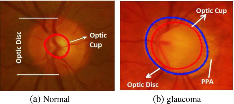
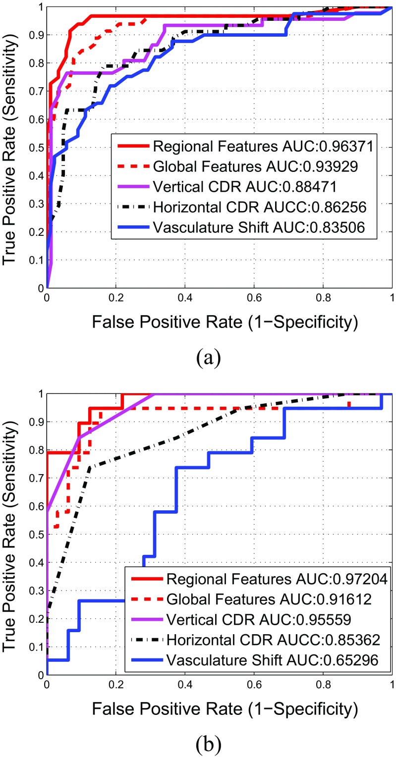
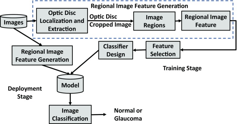
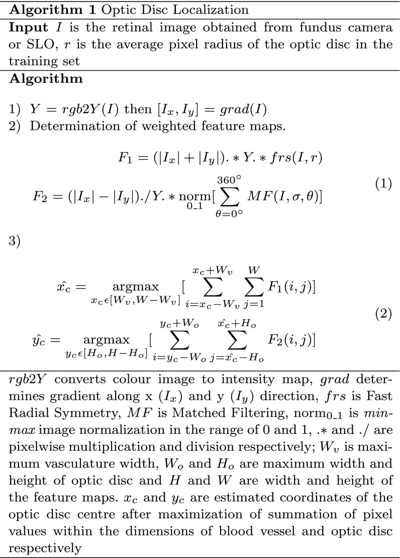
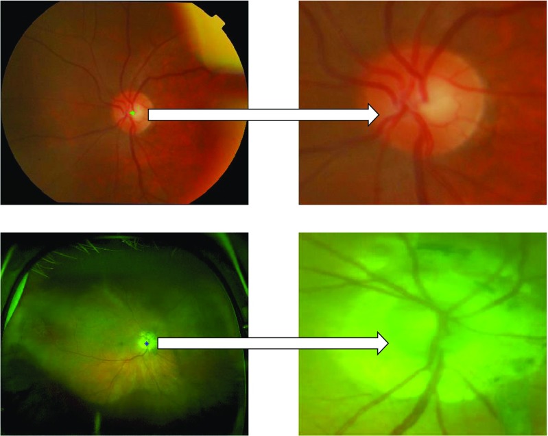
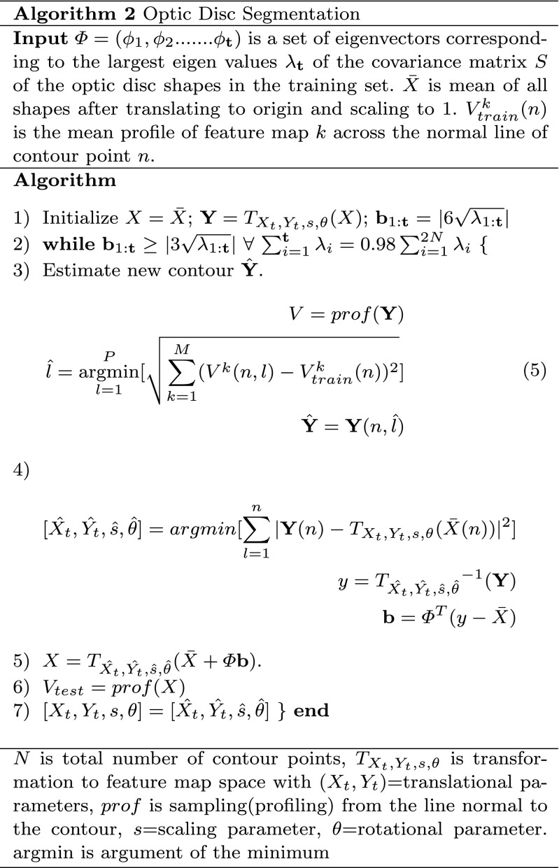
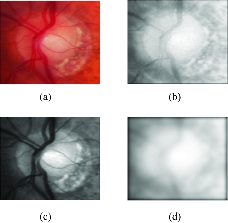
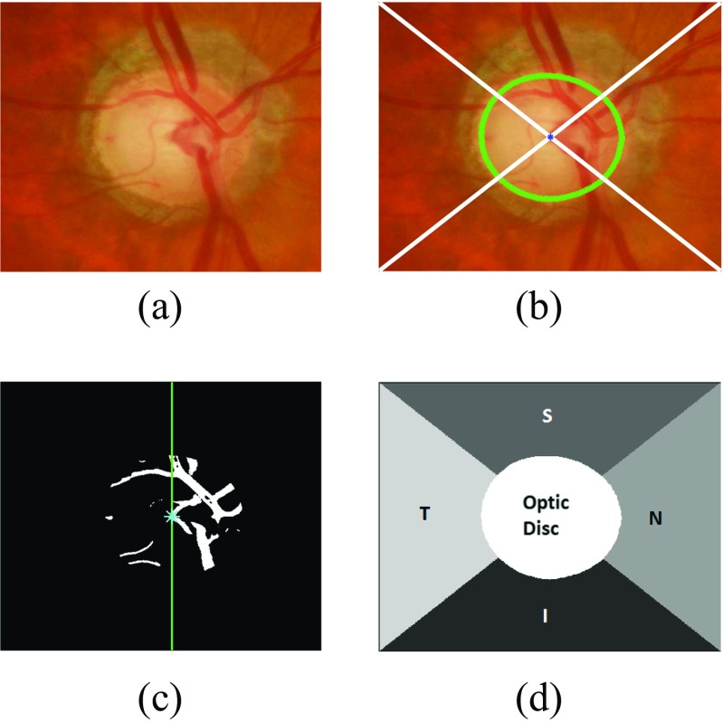
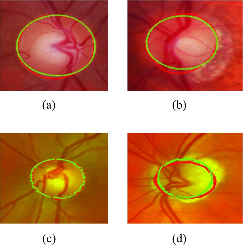
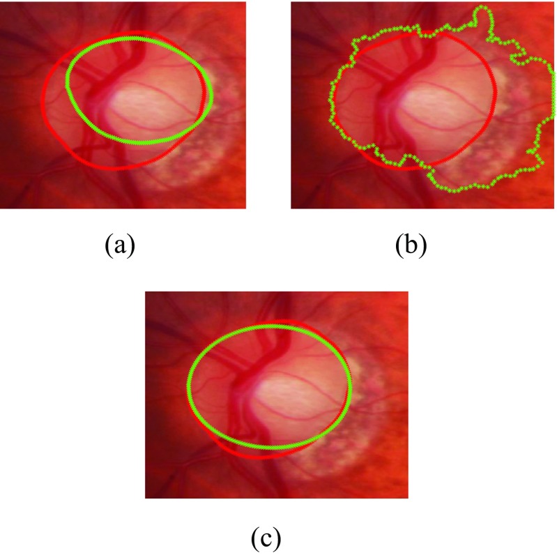
Glaucoma is one of the leading causes of blindness worldwide. There is no cure for glaucoma but detection at its earliest stage and subsequent treatment can aid patients to prevent blindness. Currently, optic disc and retinal imaging facilitates glaucoma detection but this method requires manual post-imaging modifications that are time-consuming and subjective to image assessment by human observers. Therefore, it is necessary to automate this process. In this work, we have first proposed a novel computer aided approach for automatic glaucoma detection based on Regional Image Features Model (RIFM) which can automatically perform classification between normal and glaucoma images on the basis of regional information. Different from all the existing methods, our approach can extract both geometric (e.g. morphometric properties) and non-geometric based properties (e.g. pixel appearance/intensity values, texture) from images and significantly increase the classification performance. Our proposed approach consists of three new major contributions including automatic localisation of optic disc, automatic segmentation of disc, and classification between normal and glaucoma based on geometric and non-geometric properties of different regions of an image. We have compared our method with existing approaches and tested it on both fundus and Scanning laser ophthalmoscopy (SLO) images. The experimental results show that our proposed approach outperforms the state-of-the-art approaches using either geometric or non-geometric properties. The overall glaucoma classification accuracy for fundus images is 94.4% and accuracy of detection of suspicion of glaucoma in SLO images is 93.9 %.
青光眼是全球主要的致盲原因之一。青光眼无法治愈,但在其最早阶段进行检测并随后进行治疗可帮助患者预防失明。目前,视盘和视网膜成像有助于青光眼检测,但这种方法需要在成像后进行人工修改,这既耗时,又依赖人类观察者对图像进行主观评估。因此,有必要使这个过程自动化。在这项工作中,我们首次提出了一种基于区域图像特征模型(RIFM)的新型计算机辅助自动青光眼检测方法,该方法可以根据区域信息自动对正常图像和青光眼图像进行分类。与所有现有方法不同,我们的方法可以从图像中提取几何特征(如形态测量属性)和非几何特征(如图像素外观/强度值、纹理),并显著提高分类性能。我们提出的方法包括三个新的主要贡献,即视盘的自动定位、视盘的自动分割以及基于图像不同区域的几何和非几何属性对正常和青光眼进行分类。我们将我们的方法与现有方法进行了比较,并在眼底图像和扫描激光眼底镜(SLO)图像上进行了测试。实验结果表明,我们提出的方法在使用几何或非几何属性方面均优于现有方法。眼底图像的总体青光眼分类准确率为94.4%,SLO图像中青光眼疑似病例的检测准确率为93.9%。