Honkanen Juuso T J, Turunen Mikael J, Freedman Jonathan D, Saarakkala Simo, Grinstaff Mark W, Ylärinne Janne H, Jurvelin Jukka S, Töyräs Juha
Department of Applied Physics, University of Eastern Finland, POB 1627, 70211, Kuopio, Finland.
Diagnostic Imaging Center, Kuopio University Hospital, Kuopio, Finland.
Ann Biomed Eng. 2016 Oct;44(10):2913-2921. doi: 10.1007/s10439-016-1629-z. Epub 2016 Apr 29.
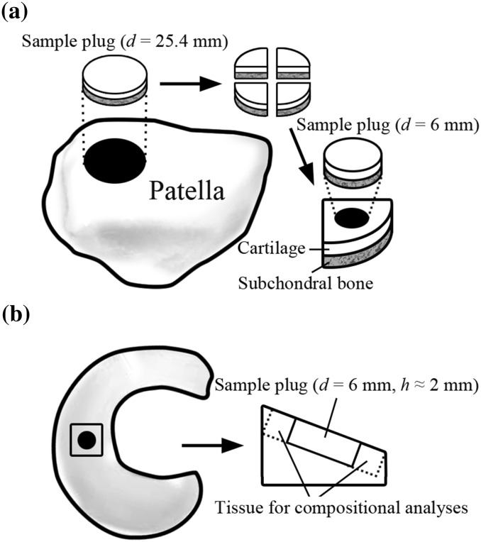
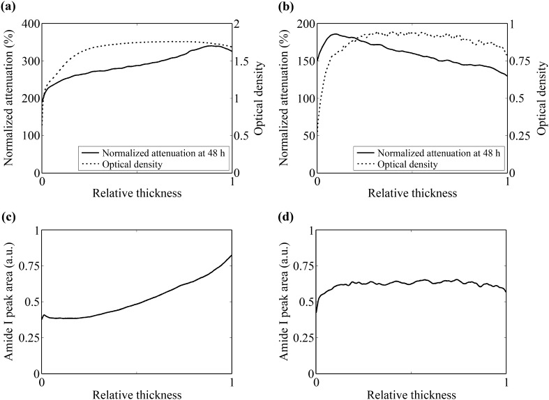
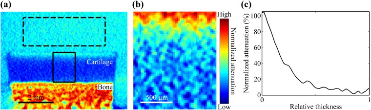
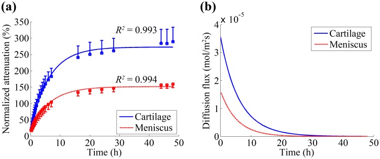
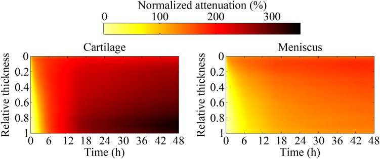
Contrast enhanced computed tomography (CECT) is a non-destructive imaging technique used for the assessment of composition and structure of articular cartilage and meniscus. Due to structural and compositional differences between these tissues, diffusion and distribution of contrast agents may differ in cartilage and meniscus. The aim of this study is to determine the diffusion kinematics of a novel iodine based cationic contrast agent (CA(2+)) in cartilage and meniscus. Cylindrical cartilage and meniscus samples (d = 6 mm, h ≈ 2 mm) were harvested from healthy bovine knee joints (n = 10), immersed in isotonic cationic contrast agent (20 mgI/mL), and imaged using a micro-CT scanner at 26 time points up to 48 h. Subsequently, normalized X-ray attenuation and contrast agent diffusion flux, as well as water, collagen and proteoglycan (PG) contents in the tissues were determined. The contrast agent distributions within cartilage and meniscus were different. In addition, the normalized attenuation and diffusion flux were higher (p < 0.05) in cartilage. Based on these results, diffusion kinematics vary between cartilage and meniscus. These tissue specific variations can affect the interpretation of CECT images and should be considered when cartilage and meniscus are assessed simultaneously.
对比增强计算机断层扫描(CECT)是一种用于评估关节软骨和半月板的组成与结构的非破坏性成像技术。由于这些组织在结构和组成上存在差异,造影剂在软骨和半月板中的扩散和分布可能有所不同。本研究的目的是确定一种新型碘基阳离子造影剂(CA(2+))在软骨和半月板中的扩散动力学。从健康牛膝关节(n = 10)获取圆柱形软骨和半月板样本(d = 6 mm,h ≈ 2 mm),将其浸入等渗阳离子造影剂(20 mgI/mL)中,并使用微型CT扫描仪在长达48小时的26个时间点进行成像。随后,测定组织中的归一化X射线衰减、造影剂扩散通量以及水、胶原蛋白和蛋白聚糖(PG)含量。造影剂在软骨和半月板中的分布不同。此外,软骨中的归一化衰减和扩散通量更高(p < 0.05)。基于这些结果,软骨和半月板之间的扩散动力学有所不同。这些组织特异性差异会影响CECT图像的解读,在同时评估软骨和半月板时应予以考虑。