• 文献检索
  • 文档翻译
  • 深度研究
  • 学术资讯
  • Suppr Zotero 插件Zotero 插件
  • 邀请有礼
  • 套餐&价格
  • 历史记录
应用&插件
Suppr Zotero 插件Zotero 插件浏览器插件Mac 客户端Windows 客户端微信小程序
定价
高级版会员购买积分包购买API积分包
服务
文献检索文档翻译深度研究API 文档MCP 服务
关于我们
关于 Suppr公司介绍联系我们用户协议隐私条款
关注我们

Suppr 超能文献

核心技术专利:CN118964589B侵权必究
粤ICP备2023148730 号-1Suppr @ 2026

文献检索

告别复杂PubMed语法,用中文像聊天一样搜索,搜遍4000万医学文献。AI智能推荐,让科研检索更轻松。

立即免费搜索

文件翻译

保留排版,准确专业,支持PDF/Word/PPT等文件格式,支持 12+语言互译。

免费翻译文档

深度研究

AI帮你快速写综述,25分钟生成高质量综述,智能提取关键信息,辅助科研写作。

立即免费体验

使用对比增强微型计算机断层扫描对半月板组织中碘摄取和放射密度进行空间和时间评估。

Spatial and temporal evaluation of iodine uptake and radiodensity in meniscus tissue using contrast-enhanced micro-CT.

作者信息

Orellana Federica, Grassi Alberto, Nuss Katja M, Wahl Peter, Neels Antonia, Zaffagnini Stefano, Parrilli Annapaola

机构信息

Empa - Swiss Federal Laboratories for Materials Science and Technology, 8600, Dübendorf, Switzerland.

Department of Chemistry, University of Fribourg, 1700, Fribourg, Switzerland.

出版信息

Heliyon. 2024 Dec 7;10(24):e41080. doi: 10.1016/j.heliyon.2024.e41080. eCollection 2024 Dec 30.

DOI:10.1016/j.heliyon.2024.e41080
PMID:39759317
原文链接:https://pmc.ncbi.nlm.nih.gov/articles/PMC11696653/
Abstract

RATIONALE AND OBJECTIVE

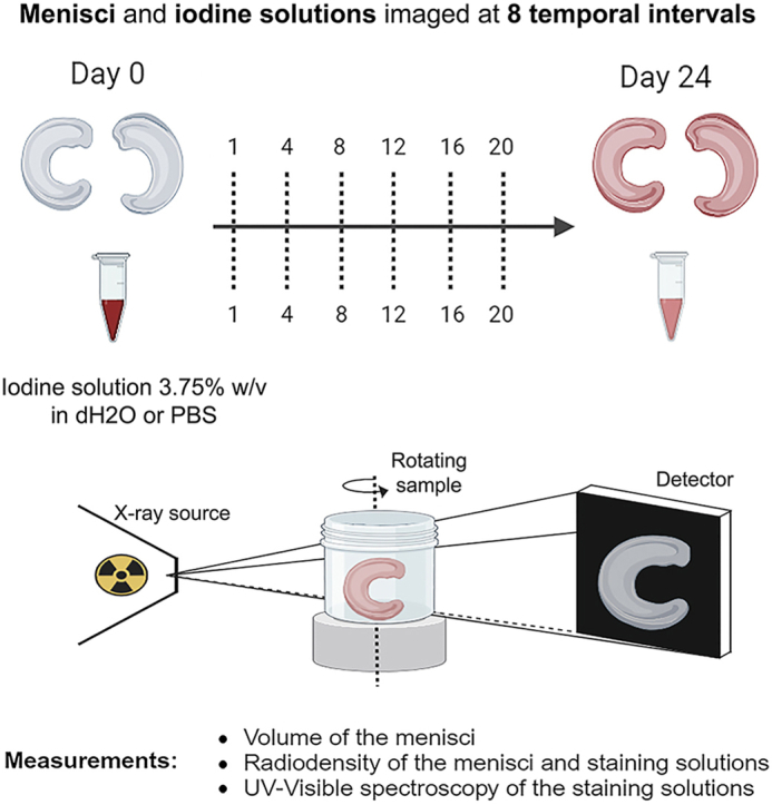
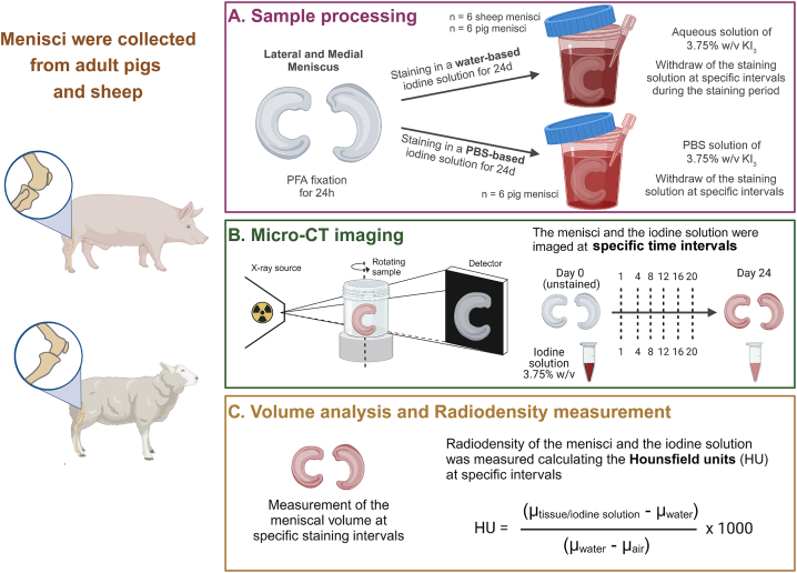
The visualization of soft tissues, like the meniscus, through X-ray micro-computed tomography (micro-CT), requires the use of contrast agents (CAs). While other studies have investigated CA diffusion in fibrocartilagineous tissues, this work aimed to optimize iodine staining protocols for meniscal tissue that improve their visualization by micro-CT. Specific objectives included evaluating the diffusion of CAs within meniscal samples over time, assessing volume changes due to staining, and identifying the iodine ions absorbed by the tissue.

MATERIALS AND METHODS

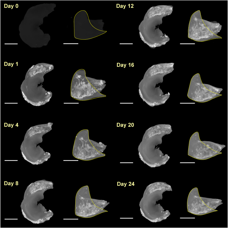
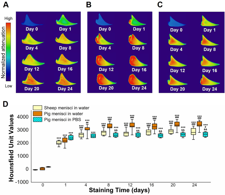
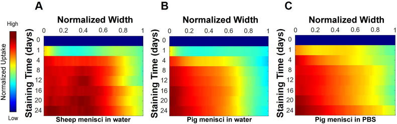
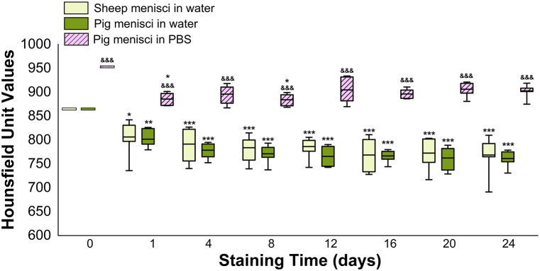
Water-based and PBS-based Lugol solutions (KI) were used to stain sheep and pig menisci for 24 days. Samples were scanned using micro-CT at different time points (0, 1, 4, 8, 12, 16, 20, and 24 days) to monitor CA diffusion and volume changes. Micro-CT provided three-dimensional (3D) visualization of iodine distribution and quantification of volume changes and radiodensity in the menisci. Additionally, UV-visible spectroscopy (UV-vis) analyses were performed to determine the uptake of iodine ions by the meniscus.

RESULTS

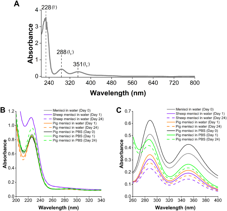
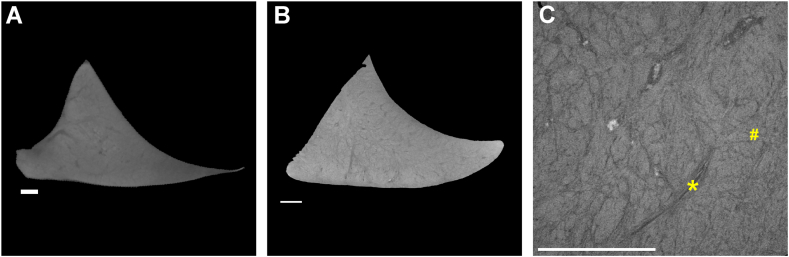
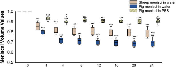
Results indicated volumetric shrinkage and increased radiodensity within the first days of staining, with diffusion primarily occurring from the periphery of the meniscus. UV-visible spectroscopy identified two iodide ions in the CA solution (I and I ) and revealed a preferential absorption of the triiodide ion (I ).

CONCLUSION

This study demonstrated the utility of iodine-based CAs and micro-CT technique for visualizing and investigating the spatial and temporal iodine diffusion within the meniscal tissue of sheep and pigs. The findings of this study have important implications for using iodine-based CAs in imaging analyses of the meniscus and offer potentially valuable insights into the diffusion patterns of iodine in fibrocartilagineous tissues.

摘要

原理与目的

通过X射线显微计算机断层扫描(显微CT)对半月板等软组织进行可视化成像,需要使用造影剂(CA)。虽然其他研究已对造影剂在纤维软骨组织中的扩散进行了调查,但本研究旨在优化半月板组织的碘染色方案,以通过显微CT改善其可视化效果。具体目标包括评估造影剂在半月板样本中的随时间扩散情况、评估染色引起的体积变化,以及确定组织吸收的碘离子。

材料与方法

使用水基和基于磷酸盐缓冲盐水(PBS)的卢戈氏溶液(KI)对绵羊和猪的半月板进行24天的染色。在不同时间点(0、1、4、8、12、16、20和24天)使用显微CT对样本进行扫描,以监测造影剂扩散和体积变化。显微CT提供了碘分布的三维(3D)可视化以及半月板体积变化和放射密度的量化。此外,还进行了紫外可见光谱(UV-vis)分析,以确定半月板对碘离子的摄取情况。

结果

结果表明,在染色的最初几天内,半月板体积缩小且放射密度增加,扩散主要发生在半月板周边。紫外可见光谱鉴定出造影剂溶液中的两种碘离子(I⁻和I₂),并显示出对三碘离子(I₃⁻)的优先吸收。

结论

本研究证明了基于碘的造影剂和显微CT技术在可视化和研究绵羊和猪半月板组织内碘的空间和时间扩散方面的实用性。本研究结果对于在半月板成像分析中使用基于碘的造影剂具有重要意义,并为碘在纤维软骨组织中的扩散模式提供了潜在的有价值见解。

https://cdn.ncbi.nlm.nih.gov/pmc/blobs/17c2/11696653/77375d74ba09/gr9.jpg
https://cdn.ncbi.nlm.nih.gov/pmc/blobs/17c2/11696653/9018b748da61/ga1.jpg
https://cdn.ncbi.nlm.nih.gov/pmc/blobs/17c2/11696653/a009790cc5fd/gr1.jpg
https://cdn.ncbi.nlm.nih.gov/pmc/blobs/17c2/11696653/f76bf01e7ccc/gr2.jpg
https://cdn.ncbi.nlm.nih.gov/pmc/blobs/17c2/11696653/de924e6312da/gr3.jpg
https://cdn.ncbi.nlm.nih.gov/pmc/blobs/17c2/11696653/28e2ee2c52b8/gr4.jpg
https://cdn.ncbi.nlm.nih.gov/pmc/blobs/17c2/11696653/a1cbb0eecd26/gr5.jpg
https://cdn.ncbi.nlm.nih.gov/pmc/blobs/17c2/11696653/e2b6a9e4fd59/gr6.jpg
https://cdn.ncbi.nlm.nih.gov/pmc/blobs/17c2/11696653/83c10b8b6771/gr7.jpg
https://cdn.ncbi.nlm.nih.gov/pmc/blobs/17c2/11696653/cb0a5522db51/gr8.jpg
https://cdn.ncbi.nlm.nih.gov/pmc/blobs/17c2/11696653/77375d74ba09/gr9.jpg

相似文献

1
Spatial and temporal evaluation of iodine uptake and radiodensity in meniscus tissue using contrast-enhanced micro-CT.使用对比增强微型计算机断层扫描对半月板组织中碘摄取和放射密度进行空间和时间评估。
Heliyon. 2024 Dec 7;10(24):e41080. doi: 10.1016/j.heliyon.2024.e41080. eCollection 2024 Dec 30.
2
Experimental assessment of diffusible iodine-based contrast-enhanced computed tomography (diceCT) protocols.基于弥散碘的对比增强计算机断层扫描(diceCT)方案的实验评估。
PeerJ. 2024 Sep 5;12:e17919. doi: 10.7717/peerj.17919. eCollection 2024.
3
Cationic Contrast Agent Diffusion Differs Between Cartilage and Meniscus.阳离子造影剂在软骨和半月板之间的扩散存在差异。
Ann Biomed Eng. 2016 Oct;44(10):2913-2921. doi: 10.1007/s10439-016-1629-z. Epub 2016 Apr 29.
4
Three-dimensional measurement of the depth of invasion in oral squamous cell carcinoma samples using Lugol's iodine-enhanced micro-computed tomography: an original study.使用卢戈氏碘增强型微型计算机断层扫描对口腔鳞状细胞癌样本侵袭深度进行三维测量:一项原创性研究。
J Appl Oral Sci. 2025 Jan 13;33:e20240304. doi: 10.1590/1678-7757-2024-0304. eCollection 2025.
5
Microstructure Analysis and Reconstruction of a Meniscus.半月板的微观结构分析与重建。
Orthop Surg. 2021 Feb;13(1):306-313. doi: 10.1111/os.12899. Epub 2021 Jan 5.
6
Iodine-Enhanced Micro-CT Imaging of Soft Tissue on the Example of Peripheral Nerve Regeneration.碘增强微 CT 成像在外周神经再生中的软组织应用。
Contrast Media Mol Imaging. 2019 Mar 27;2019:7483745. doi: 10.1155/2019/7483745. eCollection 2019.
7
Potentialities of iodine-enhanced micro-CT imaging in the morphological study of adult Aedes (Stegomyia) aegypti (Linnaeus, 1762) mosquitoes.碘增强微 CT 成像在成年埃及伊蚊(Stegomyia aegypti)形态学研究中的潜力。
Micron. 2023 Oct;173:103518. doi: 10.1016/j.micron.2023.103518. Epub 2023 Jul 21.
8
An investigation of the efficacy and mechanism of contrast-enhanced X-ray computed tomography utilizing iodine for large specimens through experimental and simulation approaches.通过实验和模拟方法对利用碘进行大样本对比增强X射线计算机断层扫描的有效性和机制进行研究。
BMC Physiol. 2015 Dec 21;15:5. doi: 10.1186/s12899-015-0019-3.
9
Iodine-enhanced micro-computed tomography of atherosclerotic plaque morphology complements conventional histology.碘增强型显微计算机断层扫描对动脉粥样硬化斑块形态的研究补充了传统组织学方法。
Atherosclerosis. 2020 Nov;313:43-49. doi: 10.1016/j.atherosclerosis.2020.09.012. Epub 2020 Sep 24.
10
Advanced soft tissue visualization in conjunction with bone structures using contrast-enhanced micro-CT.使用对比增强型显微CT结合骨结构进行高级软组织可视化。
J Med Imaging (Bellingham). 2024 Nov;11(6):066001. doi: 10.1117/1.JMI.11.6.066001. Epub 2024 Nov 22.

引用本文的文献

1
Protocol for evaluating iodine uptake and diffusion in sheep and pig meniscus tissue using contrast-enhanced micro-CT and UV-vis spectroscopy.使用对比增强型微型计算机断层扫描(micro-CT)和紫外-可见光谱法评估绵羊和猪半月板组织中碘摄取和扩散的方案。
STAR Protoc. 2025 Sep 19;6(3):103951. doi: 10.1016/j.xpro.2025.103951. Epub 2025 Jul 12.

本文引用的文献

1
Exploring contrast-enhancing staining agents for studying adipose tissue through contrast-enhanced computed tomography.探索用于通过对比增强计算机断层扫描研究脂肪组织的对比增强染色剂。
J Lipid Res. 2024 Jul;65(7):100572. doi: 10.1016/j.jlr.2024.100572. Epub 2024 May 30.
2
Revealing the complexity of meniscus microvasculature through 3D visualization and analysis.通过 3D 可视化和分析揭示半月板微血管结构的复杂性。
Sci Rep. 2024 May 13;14(1):10875. doi: 10.1038/s41598-024-61497-2.
3
Age and anatomical region-related differences in vascularization of the porcine meniscus using microcomputed tomography imaging.
使用微型计算机断层扫描成像技术对猪半月板血管化进行年龄和解剖区域相关差异研究。
J Orthop Res. 2024 Sep;42(9):2095-2105. doi: 10.1002/jor.25862. Epub 2024 Apr 29.
4
Contrast-enhanced Micro-CT 3D visualization of cell distribution in hydrated human cornea.水合人角膜中细胞分布的对比增强微型计算机断层扫描三维可视化
Heliyon. 2024 Feb 3;10(3):e25828. doi: 10.1016/j.heliyon.2024.e25828. eCollection 2024 Feb 15.
5
3D visualization of the human anterior cruciate ligament combining micro-CT and histological analysis.结合 micro-CT 和组织学分析的人体前交叉韧带的 3D 可视化。
Surg Radiol Anat. 2024 Feb;46(2):249-258. doi: 10.1007/s00276-023-03295-5. Epub 2024 Jan 24.
6
Durability assessment of hydrogel mountings for contrast-enhanced micro-CT.用于对比增强微型计算机断层扫描的水凝胶支架的耐久性评估。
Micron. 2023 Nov;174:103533. doi: 10.1016/j.micron.2023.103533. Epub 2023 Aug 29.
7
Reducing soft-tissue shrinkage artefacts caused by staining with Lugol's solution.减少卢戈氏液染色引起的软组织收缩伪影。
Sci Rep. 2021 Oct 5;11(1):19781. doi: 10.1038/s41598-021-99202-2.
8
Metabolism of vascular smooth muscle cells in vascular diseases.血管疾病中的血管平滑肌细胞代谢。
Am J Physiol Heart Circ Physiol. 2020 Sep 1;319(3):H613-H631. doi: 10.1152/ajpheart.00220.2020. Epub 2020 Aug 7.
9
Iodine-Enhanced Micro-CT Imaging of Soft Tissue on the Example of Peripheral Nerve Regeneration.碘增强微 CT 成像在外周神经再生中的软组织应用。
Contrast Media Mol Imaging. 2019 Mar 27;2019:7483745. doi: 10.1155/2019/7483745. eCollection 2019.
10
A review of artifacts in histopathology.组织病理学中的伪像综述。
J Oral Maxillofac Pathol. 2018 May-Aug;22(2):279. doi: 10.4103/jomfp.JOMFP_125_15.