Department of Periodontology, Witten/Herdecke University, Witten, Germany.
Multi-disciplinary Treatment Center, Beijing Stomatological Hospital, Capital Medical University, Beijing, China.
Clin Oral Implants Res. 2017 Jun;28(6):640-647. doi: 10.1111/clr.12848. Epub 2016 May 4.
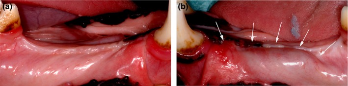
Soft tissue (ST) dehiscence with graft exposure is a frequent complication of vertical augmentation. Flap dehiscence is caused by failure to achieve tension-free primary wound closure and by the impairment of flap microcirculation due to surgical trauma. Soft tissue expansion (STE) increases ST quality and quantity prior to reconstructive surgery. We hypothesized that flap preconditioning using STE would reduce the incidence of ST complications after bone augmentation and that optimized ST healing would improve the outcome of bone regeneration.
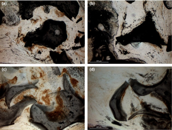
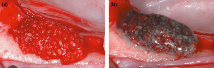
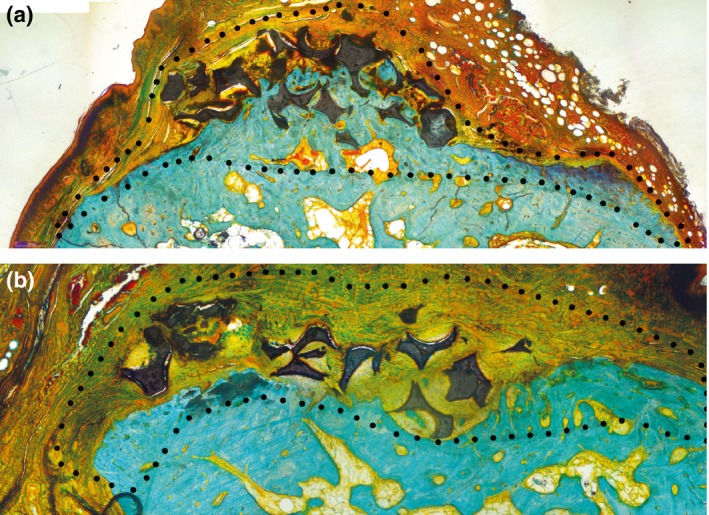
Self-filling tissue expanders were implanted in mandibular bone defects in ten beagle dogs. After expansion, alloplastic scaffolds were placed for vertical bone augmentation in STE sites and in control sites without STE pre-treatment. ST flap microcirculation was analysed using laser Doppler flowmetry. The incidence of graft exposures was evaluated after 2 weeks. Bone formation was assessed after 2 months, using histomorphometry and immunohistochemistry.
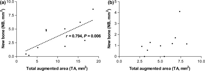
Test sites showed significantly less impairment of perfusion and faster recovery of microcirculation after bone augmentation. Furthermore, no flap dehiscences occurred in STE sites. Bone regeneration was found in both groups; however, significantly greater formation of new bone was detected in test sites with preceding STE.
Preconditioning using STE improved ST healing and bone formation after vertical augmentation. The combination of STE and the subsequent placement of alloplastic scaffolds may facilitate the reconstruction of severe bone defects.
软组织(ST)裂开伴移植物暴露是垂直增加的常见并发症。皮瓣裂开是由于未能实现无张力的一期伤口闭合和手术创伤导致皮瓣微循环受损所致。软组织扩张(STE)可增加重建手术前 ST 的质量和数量。我们假设使用 STE 对皮瓣进行预处理将减少骨增强后 ST 并发症的发生率,并且优化的 ST 愈合将改善骨再生的效果。
在 10 只比格犬的下颌骨缺损中植入自填式组织扩张器。扩张后,在 STE 部位和无 STE 预处理的对照部位放置同种异体支架进行垂直骨增强。使用激光多普勒血流仪分析 ST 皮瓣微循环。在 2 周后评估移植物暴露的发生率。在 2 个月后,通过组织形态计量学和免疫组织化学评估骨形成。
试验部位在骨增强后显示出明显较少的灌注受损和更快的微循环恢复。此外,STE 部位无皮瓣裂开。两组均发现骨再生;然而,在进行 STE 预处理的试验部位,新骨形成明显更多。
使用 STE 预处理可改善垂直增强后的 ST 愈合和骨形成。STE 与随后放置同种异体支架的结合可能有助于严重骨缺损的重建。