Long Ying, Cao Binbin, Wang Yinan, Luo Damin
School of Life Sciences, Xiamen University, Xiamen, Fujian, 361102, China.
Translational Medicine Center, Hunan Cancer Hospital, Hunan, 410006, China.
Parasit Vectors. 2016 May 17;9(1):286. doi: 10.1186/s13071-016-1568-4.
Angiostrongyliasis caused by the rat lungworm, Angiostrongylus cantonensis (A. cantonensis), has globally spread from the traditional epidemic areas. The small intestine is the site where the third-stage larvae (L3) of this worm entered the host body, and parasite proteases are involved in this process. Ac-cathB-2, a cathepsin B-like cysteine of A. cantonensis, was formerly isolated from the insoluble part of fragmentised Escherichia coli without activity. The unplanned low activity of prokaryotic expression proteins and difficulties in genetic modification hindered understanding the function of this protein.
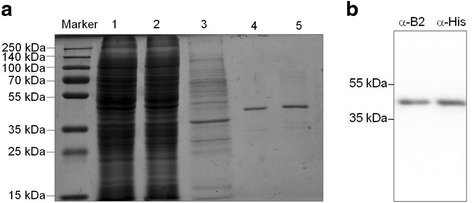
The recombinant Ac-cathB-2 was expressed and harvested from 293 T cells and the enzymatic property and the effects of processing on the activity of the recombinant protease were investigated in vitro. The expression of Ac-cathB-2 in response to external stimulation was assessed, and the function of this protease during host gut penetration was observed by using antiserum for inhibition.
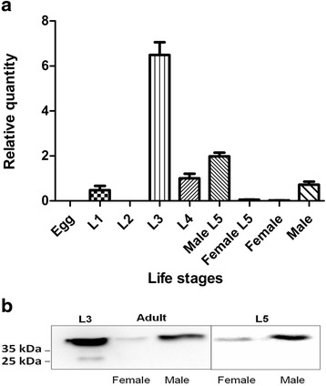
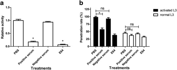
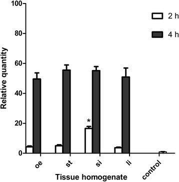
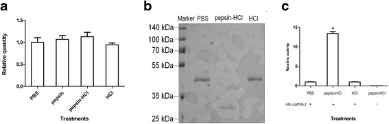
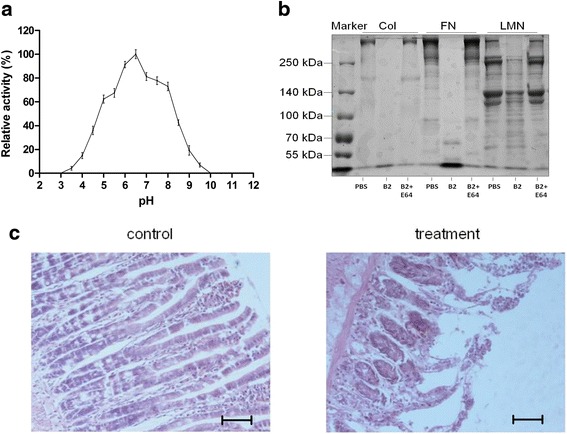
Of the life-cycle stages studied, L3 expressed the highest level of Ac-cathB-2 gene and released the corresponding gene product from the body. The expression of this gene was rapidly upregulated after incubating L3 in small intestine homogenate of rat. Recombinant Ac-cathB-2 was harvested from 293 T cell culture medium. This protease was activated by pepsin-HCl and the enabled Ac-cathB-2 could subsequently digest laminin and fibronectin readily. Moreover, the small intestine isolated from rat was disrupted after incubating with the activated Ac-cathB-2, resulting in the detachment of epithelial cells. Antiserum treatment inhibited the hydrolytic ability of recombinant Ac-cathB-2 by 82.7 %, and also reduced the tissue penetration of activated L3 by 41.2 %. Additionally, pre-incubation of L3 with artificial gastric acid increased the number of penetrating larvae by 53.2 %, and this alteration could be partly blocked by antiserum treatment.
We believe that Ac-cathB-2 from A. cantonensis might help the worm to penetrate the rat gut, because the protease was able to degrade the tissue components of host. Nevertheless, our results further indicated that host pepsin played a beneficial role in this process by cleaving Ac-cathB-2 for activation. Thus, Ac-cathB-2 may probably represent an important target for the control of A. cantonensis infection.
由广州管圆线虫(A. cantonensis)引起的管圆线虫病已从传统流行地区在全球范围内传播。小肠是该蠕虫第三期幼虫(L3)进入宿主体内的部位,寄生虫蛋白酶参与这一过程。Ac-cathB-2是广州管圆线虫的一种组织蛋白酶B样半胱氨酸蛋白酶,先前从破碎的大肠杆菌不溶性部分分离得到但无活性。原核表达蛋白意外的低活性以及基因改造的困难阻碍了对该蛋白功能的了解。
从293 T细胞中表达并收获重组Ac-cathB-2,体外研究其酶学性质以及加工处理对重组蛋白酶活性的影响。评估Ac-cathB-2对外界刺激的表达情况,并使用抗血清抑制观察该蛋白酶在宿主肠道穿透过程中的功能。
在所研究的生命周期阶段中,L3表达的Ac-cathB-2基因水平最高,并从体内释放出相应的基因产物。将L3在大鼠小肠匀浆中孵育后,该基因的表达迅速上调。从293 T细胞培养基中收获重组Ac-cathB-2。这种蛋白酶被胃蛋白酶-HCl激活,激活后的Ac-cathB-2随后能够轻易消化层粘连蛋白和纤连蛋白。此外,用激活的Ac-cathB-2孵育后,从大鼠分离的小肠被破坏,导致上皮细胞脱落。抗血清处理使重组Ac-cathB-2的水解能力降低了82.7%,并使激活的L3的组织穿透率降低了41.2%。此外,用人工胃酸预孵育L3可使穿透幼虫的数量增加53.2%,这种改变可被抗血清处理部分阻断。
我们认为广州管圆线虫的Ac-cathB-2可能有助于该蠕虫穿透大鼠肠道,因为该蛋白酶能够降解宿主的组织成分。然而,我们的结果进一步表明宿主胃蛋白酶通过切割Ac-cathB-2使其激活在这一过程中发挥了有益作用。因此,Ac-cathB-2可能是控制广州管圆线虫感染的一个重要靶点。