• 文献检索
  • 文档翻译
  • 深度研究
  • 学术资讯
  • Suppr Zotero 插件Zotero 插件
  • 邀请有礼
  • 套餐&价格
  • 历史记录
应用&插件
Suppr Zotero 插件Zotero 插件浏览器插件Mac 客户端Windows 客户端微信小程序
定价
高级版会员购买积分包购买API积分包
服务
文献检索文档翻译深度研究API 文档MCP 服务
关于我们
关于 Suppr公司介绍联系我们用户协议隐私条款
关注我们

Suppr 超能文献

核心技术专利:CN118964589B侵权必究
粤ICP备2023148730 号-1Suppr @ 2026

文献检索

告别复杂PubMed语法,用中文像聊天一样搜索,搜遍4000万医学文献。AI智能推荐,让科研检索更轻松。

立即免费搜索

文件翻译

保留排版,准确专业,支持PDF/Word/PPT等文件格式,支持 12+语言互译。

免费翻译文档

深度研究

AI帮你快速写综述,25分钟生成高质量综述,智能提取关键信息,辅助科研写作。

立即免费体验

视网膜疾病中的光学相干断层扫描血管造影术。

Optical Coherence Tomography Angiography in Retinal Diseases.

作者信息

Chalam K V, Sambhav Kumar

机构信息

Department of Ophthalmology, University of Florida College of Medicine, Florida, USA.

出版信息

J Ophthalmic Vis Res. 2016 Jan-Mar;11(1):84-92. doi: 10.4103/2008-322X.180709.

DOI:10.4103/2008-322X.180709
PMID:27195091
原文链接:https://pmc.ncbi.nlm.nih.gov/articles/PMC4860994/
Abstract

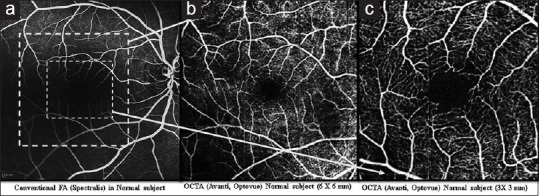
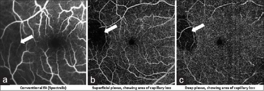
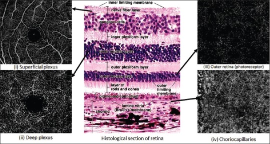
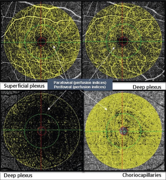
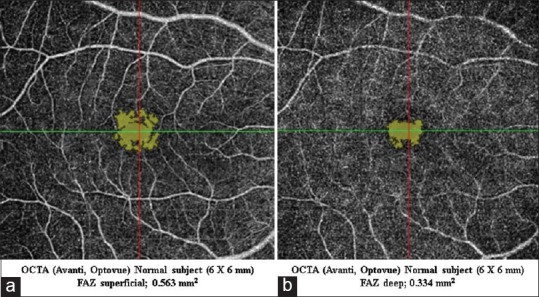
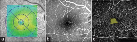
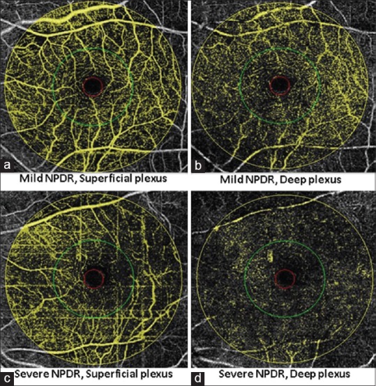
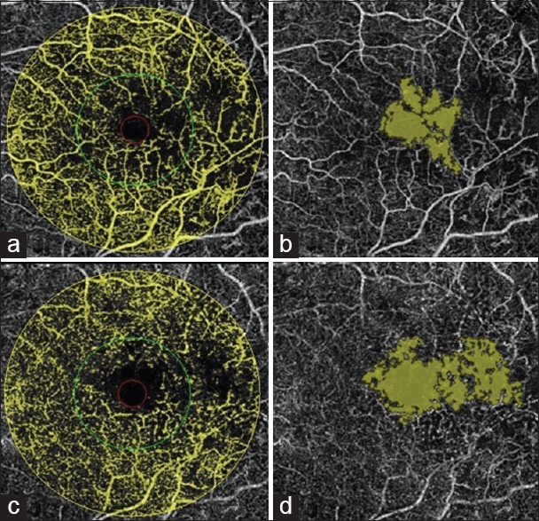
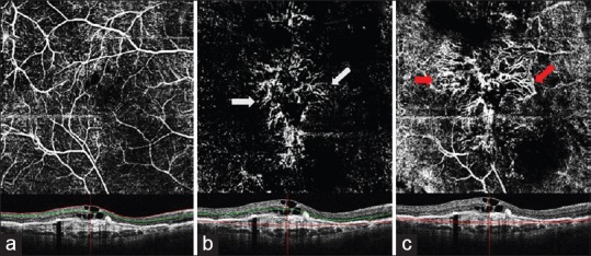
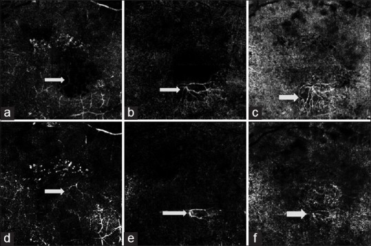
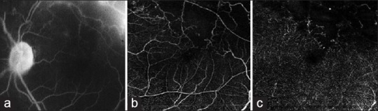
Optical coherence tomography angiography (OCTA) is a new, non-invasive imaging system that generates volumetric data of retinal and choroidal layers. It has the ability to show both structural and blood flow information. Split-spectrum amplitude-decorrelation angiography (SSADA) algorithm (a vital component of OCTA software) helps to decrease the signal to noise ratio of flow detection thus enhancing visualization of retinal vasculature using motion contrast. Published studies describe potential efficacy for OCTA in the evaluation of common ophthalmologic diseases such as diabetic retinopathy, age related macular degeneration (AMD), retinal vascular occlusions and sickle cell disease. OCTA provides a detailed view of the retinal vasculature, which allows accurate delineation of microvascular abnormalities in diabetic eyes and vascular occlusions. It helps quantify vascular compromise depending upon the severity of diabetic retinopathy. OCTA can also elucidate the presence of choroidal neovascularization (CNV) in wet AMD. In this paper, we review the knowledge, available in English language publications regarding OCTA, and compare it with the conventional angiographic standard, fluorescein angiography (FA). Finally, we summarize its potential applications to retinal vascular diseases. Its current limitations include a relatively small field of view, inability to show leakage, and tendency for image artifacts. Further larger studies will define OCTA's utility in clinical settings and establish if the technology may offer a non-invasive option of visualizing the retinal vasculature, enabling us to decrease morbidity through early detection and intervention in retinal diseases.

摘要

光学相干断层扫描血管造影(OCTA)是一种新型的非侵入性成像系统,可生成视网膜和脉络膜层的容积数据。它能够显示结构和血流信息。分裂频谱幅度去相关血管造影(SSADA)算法(OCTA软件的一个重要组成部分)有助于降低血流检测的信噪比,从而利用运动对比度增强视网膜血管系统的可视化。已发表的研究描述了OCTA在评估常见眼科疾病如糖尿病视网膜病变、年龄相关性黄斑变性(AMD)、视网膜血管阻塞和镰状细胞病方面的潜在疗效。OCTA提供了视网膜血管系统的详细视图,这使得能够准确描绘糖尿病性眼病中的微血管异常和血管阻塞情况。它有助于根据糖尿病视网膜病变的严重程度量化血管损伤。OCTA还可以阐明湿性AMD中脉络膜新生血管(CNV)的存在。在本文中,我们回顾了英文出版物中关于OCTA的知识,并将其与传统血管造影标准荧光素血管造影(FA)进行比较。最后,我们总结了其在视网膜血管疾病中的潜在应用。其目前的局限性包括视野相对较小、无法显示渗漏以及图像伪影的倾向。进一步的大型研究将确定OCTA在临床环境中的效用,并确定该技术是否可以提供一种可视化视网膜血管系统的非侵入性选择,使我们能够通过早期检测和干预视网膜疾病来降低发病率。

https://cdn.ncbi.nlm.nih.gov/pmc/blobs/7597/4860994/c02b006f936d/JOVR-11-84-g011.jpg
https://cdn.ncbi.nlm.nih.gov/pmc/blobs/7597/4860994/de5ca7383a07/JOVR-11-84-g001.jpg
https://cdn.ncbi.nlm.nih.gov/pmc/blobs/7597/4860994/65ab51921842/JOVR-11-84-g002.jpg
https://cdn.ncbi.nlm.nih.gov/pmc/blobs/7597/4860994/4c18eef11efa/JOVR-11-84-g003.jpg
https://cdn.ncbi.nlm.nih.gov/pmc/blobs/7597/4860994/b0777f411ba7/JOVR-11-84-g004.jpg
https://cdn.ncbi.nlm.nih.gov/pmc/blobs/7597/4860994/f254e8dfb38b/JOVR-11-84-g005.jpg
https://cdn.ncbi.nlm.nih.gov/pmc/blobs/7597/4860994/927cc1261be9/JOVR-11-84-g006.jpg
https://cdn.ncbi.nlm.nih.gov/pmc/blobs/7597/4860994/9b653e1abaf6/JOVR-11-84-g007.jpg
https://cdn.ncbi.nlm.nih.gov/pmc/blobs/7597/4860994/3c6b6a4d3f72/JOVR-11-84-g008.jpg
https://cdn.ncbi.nlm.nih.gov/pmc/blobs/7597/4860994/e3b7ff0e3306/JOVR-11-84-g009.jpg
https://cdn.ncbi.nlm.nih.gov/pmc/blobs/7597/4860994/76a0d48d275f/JOVR-11-84-g010.jpg
https://cdn.ncbi.nlm.nih.gov/pmc/blobs/7597/4860994/c02b006f936d/JOVR-11-84-g011.jpg

相似文献

1
Optical Coherence Tomography Angiography in Retinal Diseases.视网膜疾病中的光学相干断层扫描血管造影术。
J Ophthalmic Vis Res. 2016 Jan-Mar;11(1):84-92. doi: 10.4103/2008-322X.180709.
2
A review of optical coherence tomography angiography (OCTA).光学相干断层扫描血管造影术(OCTA)综述。
Int J Retina Vitreous. 2015 Apr 15;1:5. doi: 10.1186/s40942-015-0005-8. eCollection 2015.
3
The application of optical coherence tomography angiography in retinal diseases.光学相干断层扫描血管造影在视网膜疾病中的应用。
Surv Ophthalmol. 2017 Nov-Dec;62(6):838-866. doi: 10.1016/j.survophthal.2017.05.006. Epub 2017 Jun 1.
4
The role of optical coherence tomography angiography in fundus vascular abnormalities.光学相干断层扫描血管造影在眼底血管异常中的作用。
BMC Ophthalmol. 2016 Jul 13;16:107. doi: 10.1186/s12886-016-0277-2.
5
Classification of image artefacts in optical coherence tomography angiography of the choroid in macular diseases.黄斑疾病中脉络膜光学相干断层扫描血管造影图像伪影的分类
Clin Exp Ophthalmol. 2016 Jul;44(5):388-99. doi: 10.1111/ceo.12683. Epub 2016 Feb 11.
6
[New Possibilities in Retinal Diagnostics Using OCT Angiography].[使用光学相干断层扫描血管造影术进行视网膜诊断的新可能性]
Klin Monbl Augenheilkd. 2016 May;233(5):613-21. doi: 10.1055/s-0042-105325. Epub 2016 May 17.
7
Select Features of Diabetic Retinopathy on Swept-Source Optical Coherence Tomographic Angiography Compared With Fluorescein Angiography and Normal Eyes.比较扫频源光学相干断层血管造影与荧光素血管造影和正常眼的糖尿病视网膜病变特征。
JAMA Ophthalmol. 2016 Jun 1;134(6):644-50. doi: 10.1001/jamaophthalmol.2016.0600.
8
Optical Coherence Tomography Angiography in Eyes with Retinal Vein Occlusion.视网膜静脉阻塞患者眼部的光学相干断层扫描血管造影
J Ophthalmic Vis Res. 2018 Jul-Sep;13(3):315-332. doi: 10.4103/jovr.jovr_264_17.
9
A practical guide to optical coherence tomography angiography interpretation.光学相干断层扫描血管造影解读实用指南。
Int J Retina Vitreous. 2020 Nov 13;6(1):55. doi: 10.1186/s40942-020-00262-9.
10
Association of Choroidal Neovascularization and Central Serous Chorioretinopathy With Optical Coherence Tomography Angiography.脉络膜新生血管和中心性浆液性脉络膜视网膜病变与光学相干断层扫描血管造影的关联
JAMA Ophthalmol. 2015 Aug;133(8):899-906. doi: 10.1001/jamaophthalmol.2015.1320.

引用本文的文献

1
Assessment of retinal microvascular changes in patients with systemic lupus erythematosus using optical coherence tomography angiography.使用光学相干断层扫描血管造影术评估系统性红斑狼疮患者的视网膜微血管变化
Int J Retina Vitreous. 2025 May 8;11(1):55. doi: 10.1186/s40942-025-00677-2.
2
Effects of cigarette smoking on retinal thickness and choroidal vascularity index: a systematic review and meta-analysis.吸烟对视网膜厚度和脉络膜血管指数的影响:一项系统评价和荟萃分析。
Int J Retina Vitreous. 2025 Feb 26;11(1):21. doi: 10.1186/s40942-025-00646-9.
3
Effect of panretinal photocoagulation versus intravitreal bevacizumab injection on optic disc microcirculation in patients with diabetic retinopathy.

本文引用的文献

1
OPTICAL COHERENCE TOMOGRAPHY ANGIOGRAPHY OF TYPE 3 NEOVASCULARIZATION SECONDARY TO AGE-RELATED MACULAR DEGENERATION.年龄相关性黄斑变性继发3型新生血管的光学相干断层扫描血管造影
Retina. 2015 Nov;35(11):2229-35. doi: 10.1097/IAE.0000000000000835.
2
DETECTION OF MICROVASCULAR CHANGES IN EYES OF PATIENTS WITH DIABETES BUT NOT CLINICAL DIABETIC RETINOPATHY USING OPTICAL COHERENCE TOMOGRAPHY ANGIOGRAPHY.利用光学相干断层扫描血管造影术检测糖尿病患者但无临床糖尿病视网膜病变患者眼部的微血管变化
Retina. 2015 Nov;35(11):2364-70. doi: 10.1097/IAE.0000000000000882.
3
CORRELATION OF FOVEAL AVASCULAR ZONE SIZE WITH FOVEAL MORPHOLOGY IN NORMAL EYES USING OPTICAL COHERENCE TOMOGRAPHY ANGIOGRAPHY.
全视网膜光凝与玻璃体腔注射贝伐单抗对糖尿病视网膜病变患者视盘微循环的影响。
Int J Retina Vitreous. 2024 Dec 18;10(1):98. doi: 10.1186/s40942-024-00621-w.
4
Quantitative characterization of retinal features in translated OCTA.定量描述平移 OCTA 中的视网膜特征。
Exp Biol Med (Maywood). 2024 Oct 23;249:10333. doi: 10.3389/ebm.2024.10333. eCollection 2024.
5
Evaluation of Foveal Vasculature by Optical Coherence Tomography Angiography after Pan-Retinal Photocoagulation versus Intravitreal Anti-VEGF Injections.全视网膜光凝与玻璃体腔内抗血管内皮生长因子注射后光学相干断层扫描血管造影对黄斑区血管系统的评估
J Ophthalmic Vis Res. 2024 Sep 16;19(3):313-323. doi: 10.18502/jovr.v19i3.13622. eCollection 2024 Jul-Sep.
6
Research progress of optic nerve imaging during 1991-2023: a bibliometric analysis.1991 - 2023年视神经成像的研究进展:一项文献计量分析
Quant Imaging Med Surg. 2024 Sep 1;14(9):6566-6578. doi: 10.21037/qims-24-870. Epub 2024 Aug 28.
7
Optical coherence tomography angiography in neovascular age-related macular degeneration: comprehensive review of advancements and future perspective.光学相干断层扫描血管造影在新生血管性年龄相关性黄斑变性中的应用:进展与未来展望的综合综述
Eye (Lond). 2025 Apr;39(5):835-844. doi: 10.1038/s41433-024-03295-8. Epub 2024 Aug 15.
8
Soul: An OCTA dataset based on Human Machine Collaborative Annotation Framework.灵魂:基于人机协作标注框架的 OCTA 数据集。
Sci Data. 2024 Aug 2;11(1):838. doi: 10.1038/s41597-024-03665-7.
9
Role of optical coherence tomography angiography in retinal tumors: A narrative review.光学相干断层扫描血管造影在视网膜肿瘤中的作用:叙述性综述。
Indian J Ophthalmol. 2024 Aug 1;72(8):1082-1090. doi: 10.4103/IJO.IJO_29_24. Epub 2024 Jul 29.
10
Optical coherence tomography angiography evaluation of retinal and choroidal microvascular changes in pregnant women with a history of unexplained recurrent spontaneous abortions.光学相干断层扫描血管造影术评估有不明原因复发性自然流产史孕妇的视网膜和脉络膜微血管变化
Arch Gynecol Obstet. 2024 Aug;310(2):883-892. doi: 10.1007/s00404-024-07549-4. Epub 2024 May 30.
使用光学相干断层扫描血管造影术对正常眼黄斑中心凹血管区大小与黄斑中心凹形态的相关性研究
Retina. 2015 Nov;35(11):2188-95. doi: 10.1097/IAE.0000000000000847.
4
DETECTION OF NONEXUDATIVE CHOROIDAL NEOVASCULARIZATION IN AGE-RELATED MACULAR DEGENERATION WITH OPTICAL COHERENCE TOMOGRAPHY ANGIOGRAPHY.光学相干断层扫描血管造影术检测年龄相关性黄斑变性中的非渗出性脉络膜新生血管
Retina. 2015 Nov;35(11):2204-11. doi: 10.1097/IAE.0000000000000867.
5
ASSOCIATION BETWEEN CHANGES IN MACULAR VASCULATURE IN OPTICAL COHERENCE TOMOGRAPHY- AND FLUORESCEIN- ANGIOGRAPHY AND DISTRIBUTION OF MACULAR PIGMENT IN TYPE 2 IDIOPATHIC MACULAR TELANGIECTASIA.光学相干断层扫描和荧光素血管造影中黄斑血管变化与2型特发性黄斑毛细血管扩张症中黄斑色素分布的关联
Retina. 2015 Nov;35(11):2307-16. doi: 10.1097/IAE.0000000000000868.
6
RETINAL VASCULAR PERFUSION DENSITY MAPPING USING OPTICAL COHERENCE TOMOGRAPHY ANGIOGRAPHY IN NORMALS AND DIABETIC RETINOPATHY PATIENTS.使用光学相干断层扫描血管造影术对正常人和糖尿病视网膜病变患者进行视网膜血管灌注密度映射
Retina. 2015 Nov;35(11):2353-63. doi: 10.1097/IAE.0000000000000862.
7
LONGITUDINAL OPTICAL COHERENCE TOMOGRAPHY-ANGIOGRAPHY STUDY OF TYPE 2 NAIVE CHOROIDAL NEOVASCULARIZATION EARLY RESPONSE AFTER TREATMENT.2型初发性脉络膜新生血管治疗后早期反应的纵向光学相干断层扫描血管造影研究
Retina. 2015 Nov;35(11):2242-51. doi: 10.1097/IAE.0000000000000879.
8
OPTICAL COHERENCE TOMOGRAPHY ANGIOGRAPHY IN EARLY TYPE 3 NEOVASCULARIZATION.光学相干断层扫描血管造影术在早期3型新生血管形成中的应用
Retina. 2015 Nov;35(11):2236-41. doi: 10.1097/IAE.0000000000000834.
9
OPTICAL COHERENCE TOMOGRAPHY ANGIOGRAPHY IN RETINAL ARTERY OCCLUSION.视网膜动脉阻塞的光学相干断层扫描血管造影术
Retina. 2015 Nov;35(11):2339-46. doi: 10.1097/IAE.0000000000000850.
10
ENLARGEMENT OF FOVEAL AVASCULAR ZONE IN DIABETIC EYES EVALUATED BY EN FACE OPTICAL COHERENCE TOMOGRAPHY ANGIOGRAPHY.通过正面光学相干断层扫描血管造影评估糖尿病患者眼部黄斑血管区扩大情况。
Retina. 2015 Nov;35(11):2377-83. doi: 10.1097/IAE.0000000000000849.