Qu Xuefeng, Yan Jiaqing, Li Xiaoli, Zhang Peixun, Liu Xianzeng
Division of the Comprehensive Epilepsy Center and Neurofunctional Monitoring Laboratory, Department of Neurology, Peking University People's Hospital Beijing, China.
School of Electrical and Control Engineering, North China University of Technology Beijing, China.
Front Comput Neurosci. 2016 May 4;10:43. doi: 10.3389/fncom.2016.00043. eCollection 2016.
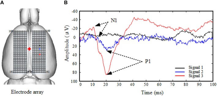
Traditionally, the topography of somatosensory evoked potentials (SEPs) is generated based on amplitude and latency. However, this operation focuses on the physical morphology and field potential-power, so it suffers from difficulties in performing identification in an objective manner. In this study, measurement of the synchronization of SEPs is proposed as a method to explore brain functional networks as well as the plasticity after peripheral nerve injury.
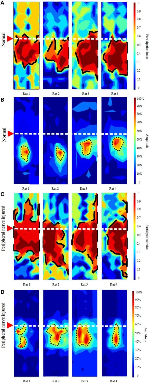
SEPs elicited by unilateral sciatic nerve stimulation in twelve adult male Sprague-Dawley (SD) rats in the normal group were compared with SEPs evoked after unilateral sciatic nerve hemisection in four peripheral nerve injured SD rats. The characterization of synchronized networks from SEPs was conducted using equal-time correlation, correlation matrix analysis, and comparison to randomized surrogate data. Eigenvalues of the correlation matrix were used to identify the clusters of functionally synchronized neuronal activity, and the participation index (PI) was calculated to indicate the involvement of each channel in the cluster. The PI value at the knee point of the PI histogram was used as a threshold to demarcate the cortical boundary.
Ten out of the twelve normal rats showed only one synchronized brain network. The remaining two normal rats showed one strong and one weak network. In the peripheral nerve injured group, only one synchronized brain network was found in each rat. In the normal group, all network shapes appear regular and the network is largely contained in the posterior cortex. In the injured group, the network shapes appear irregular, the network extends anteriorly and posteriorly, and the network area is significantly larger. There are considerable individual variations in the shape and location of the network after peripheral nerve injury.
The proposed method can detect functional brain networks. Compared to the results of the traditional SEP-morphology-based analysis method, the synchronized functional network area is much larger. Furthermore, the proposed method can also characterize the rapid cortical plasticity after a peripheral nerve is acutely injured.
传统上,体感诱发电位(SEP)的地形图是基于振幅和潜伏期生成的。然而,这种操作侧重于物理形态和场电位功率,因此在进行客观识别时存在困难。在本研究中,提出测量SEP的同步性,作为探索脑功能网络以及周围神经损伤后可塑性的一种方法。
将正常组12只成年雄性Sprague-Dawley(SD)大鼠单侧坐骨神经刺激诱发的SEP与4只周围神经损伤的SD大鼠单侧坐骨神经半切术后诱发的SEP进行比较。使用等时相关性、相关矩阵分析以及与随机替代数据的比较,对SEP同步网络的特征进行分析。相关矩阵的特征值用于识别功能同步神经元活动的簇,并计算参与指数(PI)以表明每个通道在簇中的参与情况。PI直方图拐点处的PI值用作划分皮质边界的阈值。
12只正常大鼠中有10只仅显示一个同步脑网络。其余2只正常大鼠显示一个强网络和一个弱网络。在周围神经损伤组中,每只大鼠仅发现一个同步脑网络。在正常组中,所有网络形状均规则,且网络主要包含在后皮质中。在损伤组中,网络形状不规则,网络向前和向后延伸,且网络面积明显更大。周围神经损伤后,网络的形状和位置存在相当大的个体差异。
所提出的方法可以检测脑功能网络。与传统的基于SEP形态学分析方法的结果相比,同步功能网络面积要大得多。此外,所提出的方法还可以表征周围神经急性损伤后快速的皮质可塑性。