• 文献检索
  • 文档翻译
  • 深度研究
  • 学术资讯
  • Suppr Zotero 插件Zotero 插件
  • 邀请有礼
  • 套餐&价格
  • 历史记录
应用&插件
Suppr Zotero 插件Zotero 插件浏览器插件Mac 客户端Windows 客户端微信小程序
定价
高级版会员购买积分包购买API积分包
服务
文献检索文档翻译深度研究API 文档MCP 服务
关于我们
关于 Suppr公司介绍联系我们用户协议隐私条款
关注我们

Suppr 超能文献

核心技术专利:CN118964589B侵权必究
粤ICP备2023148730 号-1Suppr @ 2026

文献检索

告别复杂PubMed语法,用中文像聊天一样搜索,搜遍4000万医学文献。AI智能推荐,让科研检索更轻松。

立即免费搜索

文件翻译

保留排版,准确专业,支持PDF/Word/PPT等文件格式,支持 12+语言互译。

免费翻译文档

深度研究

AI帮你快速写综述,25分钟生成高质量综述,智能提取关键信息,辅助科研写作。

立即免费体验

软骨下骨结构与骨关节炎组织病理学分级之间的关联。

Association between subchondral bone structure and osteoarthritis histopathological grade.

作者信息

Finnilä Mikko A J, Thevenot Jérôme, Aho Olli-Matti, Tiitu Virpi, Rautiainen Jari, Kauppinen Sami, Nieminen Miika T, Pritzker Kenneth, Valkealahti Maarit, Lehenkari Petri, Saarakkala Simo

机构信息

Research Unit of Medical Imaging, Physics and Technology, Faculty of Medicine, University of Oulu, Oulu, Finland.

Medical Research Center Oulu, Oulu University Hospital and University of Oulu, Oulu, Finland.

出版信息

J Orthop Res. 2017 Apr;35(4):785-792. doi: 10.1002/jor.23312. Epub 2016 Jun 22.

DOI:10.1002/jor.23312
PMID:27227565
原文链接:https://pmc.ncbi.nlm.nih.gov/articles/PMC5412847/
Abstract

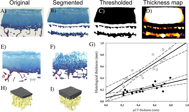
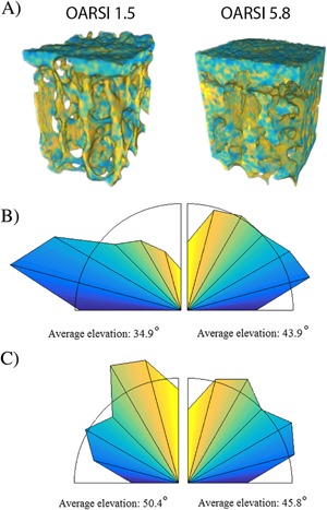
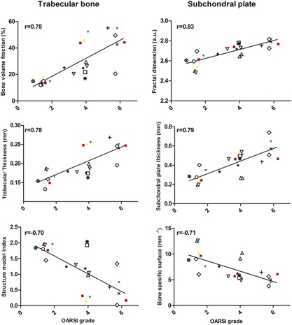
Despite increasing evidence that subchondral bone contributes to osteoarthritis (OA) pathogenesis, little is known about local changes in bone structure compared to cartilage degeneration. This study linked structural adaptation of subchondral bone with histological OA grade. Twenty-five osteochondral samples of macroscopically different degeneration were prepared from tibiae of 14 patients. Samples were scanned with micro-computed tomography (μCT) and both conventional structural parameters and novel 3D parameters based on local patterns were analyzed from the subchondral plate and trabecular bone. Subsequently, samples were processed for histology and evaluated for OARSI grade. Each bone parameter and OARSI grade was compared to assess structural adaptation of bone with OA severity. In addition, thicknesses of cartilage, calcified cartilage, and subchondral plate were analyzed from histological sections and compared with subchondral bone plate thickness from μCT. With increasing OARSI grade, the subchondral plate became thicker along with decreased specific bone surface, while there was no change in tissue mineral density. Histological analysis showed that subchondral plate thickness from μCT also includes calcified cartilage. Entropy of local patterns increased with OA severity, reflecting higher tissue heterogeneity. In the trabecular compartment, bone volume fraction and both trabecular thickness and number increased with OARSI grade while trabecular separation and structure model index decreased. Also, elevation of local patterns became longitudinal in the subchondral plate and axial transverse in trabecular bone with increasing OARSI grade. This study demonstrates the possibility of radiological assessment of OA severity by structural analysis of bone. © 2016 The Authors. Journal of Orthopaedic Research Published by Wiley Periodicals, Inc. J Orthop Res 35:785-792, 2017.

摘要

尽管越来越多的证据表明软骨下骨在骨关节炎(OA)发病机制中起作用,但与软骨退变相比,关于骨结构的局部变化却知之甚少。本研究将软骨下骨的结构适应性与组织学OA分级联系起来。从14例患者的胫骨制备了25个宏观退变程度不同的骨软骨样本。用微计算机断层扫描(μCT)对样本进行扫描,并分析软骨下板和松质骨的传统结构参数以及基于局部模式的新型三维参数。随后,对样本进行组织学处理并评估OARSI分级。比较每个骨参数和OARSI分级,以评估骨的结构适应性与OA严重程度的关系。此外,从组织学切片分析软骨、钙化软骨和软骨下板的厚度,并与μCT测量的软骨下骨板厚度进行比较。随着OARSI分级增加,软骨下板变厚,同时比骨表面积减小,而组织矿物质密度无变化。组织学分析表明,μCT测量的软骨下板厚度也包括钙化软骨。局部模式的熵随OA严重程度增加,反映出更高的组织异质性。在松质骨区域,骨体积分数、小梁厚度和数量随OARSI分级增加,而小梁间距和结构模型指数减小。而且,随着OARSI分级增加,局部模式的升高在软骨下板变为纵向,在松质骨变为轴向横向。本研究证明了通过骨结构分析对OA严重程度进行放射学评估的可能性。© 2016作者。《骨科研究杂志》由威利期刊公司出版。《骨科研究杂志》35:785 - 792,2017年。

https://cdn.ncbi.nlm.nih.gov/pmc/blobs/0875/5412847/33771e30e50b/JOR-35-785-g003.jpg
https://cdn.ncbi.nlm.nih.gov/pmc/blobs/0875/5412847/977a7ccbfeb3/JOR-35-785-g001.jpg
https://cdn.ncbi.nlm.nih.gov/pmc/blobs/0875/5412847/b4187e45ba51/JOR-35-785-g002.jpg
https://cdn.ncbi.nlm.nih.gov/pmc/blobs/0875/5412847/33771e30e50b/JOR-35-785-g003.jpg
https://cdn.ncbi.nlm.nih.gov/pmc/blobs/0875/5412847/977a7ccbfeb3/JOR-35-785-g001.jpg
https://cdn.ncbi.nlm.nih.gov/pmc/blobs/0875/5412847/b4187e45ba51/JOR-35-785-g002.jpg
https://cdn.ncbi.nlm.nih.gov/pmc/blobs/0875/5412847/33771e30e50b/JOR-35-785-g003.jpg

相似文献

1
Association between subchondral bone structure and osteoarthritis histopathological grade.软骨下骨结构与骨关节炎组织病理学分级之间的关联。
J Orthop Res. 2017 Apr;35(4):785-792. doi: 10.1002/jor.23312. Epub 2016 Jun 22.
2
Correlation between subchondral bone plate thickness and cartilage degeneration in osteoarthritis of the ankle.踝关节骨关节炎中软骨下骨板厚度与软骨退变的相关性
Foot Ankle Int. 2014 Dec;35(12):1341-9. doi: 10.1177/1071100714548061. Epub 2014 Aug 18.
3
Subchondral bone histology and grading in osteoarthritis.骨关节炎中的软骨下骨组织学及分级
PLoS One. 2017 Mar 20;12(3):e0173726. doi: 10.1371/journal.pone.0173726. eCollection 2017.
4
Spatial and temporal changes of subchondral bone proceed to microscopic articular cartilage degeneration in guinea pigs with spontaneous osteoarthritis.自发性骨关节炎豚鼠的软骨下骨时空变化先于微观关节软骨退变。
Osteoarthritis Cartilage. 2013 Apr;21(4):574-81. doi: 10.1016/j.joca.2013.01.002. Epub 2013 Jan 9.
5
Application of in vivo micro-computed tomography in the temporal characterisation of subchondral bone architecture in a rat model of low-dose monosodium iodoacetate-induced osteoarthritis.体内 micro-CT 在低剂量碘酸钠诱导的骨关节炎大鼠模型中关节软骨下骨结构的时相特征研究中的应用。
Arthritis Res Ther. 2011;13(6):R210. doi: 10.1186/ar3543. Epub 2011 Dec 21.
6
Parathyroid hormone (1-34) prevents cartilage degradation and preserves subchondral bone micro-architecture in guinea pigs with spontaneous osteoarthritis.甲状旁腺激素(1-34)可预防自发性骨关节炎豚鼠的软骨降解并保留软骨下骨微结构。
Osteoarthritis Cartilage. 2014 Nov;22(11):1869-77. doi: 10.1016/j.joca.2014.07.013. Epub 2014 Jul 30.
7
Bone marrow lesions in knee osteoarthritis: regional differences in tibial subchondral bone microstructure and their association with cartilage degeneration.膝关节骨关节炎骨髓病变:胫骨软骨下骨微观结构的区域性差异及其与软骨退变的关系。
Osteoarthritis Cartilage. 2019 Nov;27(11):1653-1662. doi: 10.1016/j.joca.2019.07.004. Epub 2019 Jul 12.
8
Correlation between 3D microstructural and 2D histomorphometric properties of subchondral bone with healthy and degenerative cartilage of the knee joint.膝关节健康软骨和退变软骨下骨的三维微观结构与二维组织形态计量学特性之间的相关性
Histol Histopathol. 2014 Nov;29(11):1477-88. doi: 10.14670/HH-29.1477. Epub 2014 May 14.
9
3D morphometric analysis of calcified cartilage properties using micro-computed tomography.使用微计算机断层扫描对钙化软骨特性进行 3D 形态计量分析。
Osteoarthritis Cartilage. 2019 Jan;27(1):172-180. doi: 10.1016/j.joca.2018.09.009. Epub 2018 Oct 1.
10
Bone turnover and articular cartilage differences localized to subchondral cysts in knees with advanced osteoarthritis.骨转换和关节软骨差异定位于晚期骨关节炎膝关节的软骨下囊肿。
Osteoarthritis Cartilage. 2015 Dec;23(12):2174-2183. doi: 10.1016/j.joca.2015.07.012. Epub 2015 Aug 1.

引用本文的文献

1
Research Progress of Osteoarthritis Treatment by Low Intensity Pulsed Ultrasound.低强度脉冲超声治疗骨关节炎的研究进展
Smart Med. 2025 May 16;4(2):e70003. doi: 10.1002/smmd.70003. eCollection 2025 Jun.
2
Changes in subchondral bone morphology with osteoarthritis in the ankle.踝关节骨关节炎的软骨下骨形态变化。
PLoS One. 2024 Jun 18;19(6):e0290914. doi: 10.1371/journal.pone.0290914. eCollection 2024.
3
Biomechanics of the Human Osteochondral Unit: A Systematic Review.人体骨软骨单元的生物力学:系统评价

本文引用的文献

1
Alterations in subchondral bone plate, trabecular bone and articular cartilage properties of rabbit femoral condyles at 4 weeks after anterior cruciate ligament transection.前交叉韧带横断术后4周兔股骨髁软骨下骨板、松质骨及关节软骨特性的改变
Osteoarthritis Cartilage. 2015 Mar;23(3):414-22. doi: 10.1016/j.joca.2014.11.023. Epub 2014 Dec 3.
2
Quantification of differences in bone texture from plain radiographs in knees with and without osteoarthritis.对患骨关节炎和未患骨关节炎膝关节的X线平片上骨质纹理差异的量化分析。
Osteoarthritis Cartilage. 2014 Oct;22(10):1724-31. doi: 10.1016/j.joca.2014.06.021.
3
Subchondral bone in osteoarthritis: insight into risk factors and microstructural changes.
Materials (Basel). 2024 Apr 8;17(7):1698. doi: 10.3390/ma17071698.
4
Clinical Super-Resolution Computed Tomography of Bone Microstructure: Application in Musculoskeletal and Dental Imaging.临床超分辨率计算机断层扫描骨微结构:肌肉骨骼和牙科成像中的应用。
Ann Biomed Eng. 2024 May;52(5):1255-1269. doi: 10.1007/s10439-024-03450-y. Epub 2024 Feb 15.
5
Subchondral bone remodeling patterns in larger animal models of meniscal injuries inducing knee osteoarthritis - a systematic review.半月板损伤致膝骨关节炎较大动物模型的软骨下骨重塑模式:系统评价。
Knee Surg Sports Traumatol Arthrosc. 2023 Dec;31(12):5346-5364. doi: 10.1007/s00167-023-07579-6. Epub 2023 Sep 24.
6
Exercise improves subchondral bone microenvironment through regulating bone-cartilage crosstalk.运动通过调节骨-软骨串扰改善软骨下骨微环境。
Front Endocrinol (Lausanne). 2023 May 23;14:1159393. doi: 10.3389/fendo.2023.1159393. eCollection 2023.
7
The effect of postmenopausal osteoporosis on subchondral bone pathology in a rat model of knee osteoarthritis.绝经后骨质疏松症对膝骨关节炎大鼠模型软骨下骨病理学的影响。
Sci Rep. 2023 Feb 20;13(1):2926. doi: 10.1038/s41598-023-29802-7.
8
3D profiling of mouse epiphyses across ages reveals new potential imaging biomarkers of early spontaneous osteoarthritis.对不同年龄小鼠干骺端的 3D 分析揭示了早期自发性骨关节炎新的潜在影像学生物标志物。
J Anat. 2023 Jun;242(6):1037-1050. doi: 10.1111/joa.13834. Epub 2023 Feb 11.
9
Morphological and mechanical alterations in articular cartilage and subchondral bone during spontaneous hip osteoarthritis in guinea pigs.豚鼠自发性髋骨关节炎期间关节软骨和软骨下骨的形态学和力学改变
Front Bioeng Biotechnol. 2023 Jan 23;11:1080241. doi: 10.3389/fbioe.2023.1080241. eCollection 2023.
10
Novel micro-MRI approach for subchondral trabecular bone analysis in patients with hip osteoarthritis is comparable to micro-CT approach.新型微磁共振成像方法可用于髋关节骨关节炎患者的软骨下骨小梁分析,与微计算机断层扫描方法相当。
Croat Med J. 2022 Dec 31;63(6):515-524. doi: 10.3325/cmj.2022.63.515.
骨关节炎中的软骨下骨:对危险因素和微观结构变化的洞察
Arthritis Res Ther. 2013;15(6):223. doi: 10.1186/ar4405.
4
Osteoarthritis--a case for personalized health care?骨关节炎——个性化医疗保健的一个案例?
Osteoarthritis Cartilage. 2014 Jan;22(1):7-16. doi: 10.1016/j.joca.2013.10.018. Epub 2013 Nov 8.
5
Inhibition of TGF-β signaling in mesenchymal stem cells of subchondral bone attenuates osteoarthritis.抑制软骨下骨间充质干细胞中的 TGF-β 信号通路可减轻骨关节炎。
Nat Med. 2013 Jun;19(6):704-12. doi: 10.1038/nm.3143. Epub 2013 May 19.
6
Spatial and temporal changes of subchondral bone proceed to microscopic articular cartilage degeneration in guinea pigs with spontaneous osteoarthritis.自发性骨关节炎豚鼠的软骨下骨时空变化先于微观关节软骨退变。
Osteoarthritis Cartilage. 2013 Apr;21(4):574-81. doi: 10.1016/j.joca.2013.01.002. Epub 2013 Jan 9.
7
Direct assessment of articular cartilage and underlying subchondral bone reveals a progressive gene expression change in human osteoarthritic knees.直接评估关节软骨和其下的软骨下骨揭示了人类骨关节炎膝关节中进行性的基因表达变化。
Osteoarthritis Cartilage. 2013 Mar;21(3):450-61. doi: 10.1016/j.joca.2012.11.016. Epub 2012 Dec 5.
8
Enhanced trabecular micro-architecture of the femoral neck in hip osteoarthritis vs. healthy controls: a micro-computer tomography study in postmenopausal women.髋关节骨关节炎与健康对照组患者股骨颈小梁微结构的增强:绝经后女性的微计算机断层扫描研究。
Int Orthop. 2013 Jan;37(1):21-6. doi: 10.1007/s00264-012-1713-4. Epub 2012 Nov 24.
9
Alterations to the subchondral bone architecture during osteoarthritis: bone adaptation vs endochondral bone formation.骨关节炎过程中软骨下骨结构的改变:骨适应性与软骨内成骨。
Osteoarthritis Cartilage. 2013 Feb;21(2):331-8. doi: 10.1016/j.joca.2012.10.013. Epub 2012 Nov 7.
10
Difference in subchondral cancellous bone between postmenopausal women with hip osteoarthritis and osteoporotic fracture: implication for fatigue microdamage, bone microarchitecture, and biomechanical properties.绝经后髋骨关节炎女性与骨质疏松性骨折女性的软骨下松质骨差异:对疲劳微损伤、骨微结构和生物力学性能的影响
Arthritis Rheum. 2012 Dec;64(12):3955-62. doi: 10.1002/art.34670.