Feke Gilbert T, Hyman Bradley T, Stern Robert A, Pasquale Louis R
Department of Ophthalmology, Massachusetts Eye and Ear Infirmary, Harvard Medical, School, Boston, MA, USA.
Department of Neurology, Massachusetts Alzheimer's Disease Research Center, Massachusetts General Hospital, Harvard Medical School, Boston, MA, USA.
Alzheimers Dement (Amst). 2015 Apr 23;1(2):144-51. doi: 10.1016/j.dadm.2015.01.004. eCollection 2015 Jun.
Patients with Alzheimer's disease (AD) demonstrate the narrowing of retinal veins and decreased retinal venous blood flow compared with control subjects. We assessed whether these abnormalities are present in patients with mild cognitive impairment (MCI).
After the determination of the global clinical dementia rating, 52 subjects (10 AD, 21 MCI, and 21 normal controls) underwent retinal hemodynamic profiling. Blood column diameter, blood speed, and blood flow were measured in a major temporal retinal vein using retinal laser Doppler flowmetry. In addition, peripapillary retinal nerve fiber layer (RNFL) thickness was measured using optical coherence tomography.
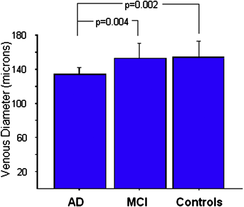
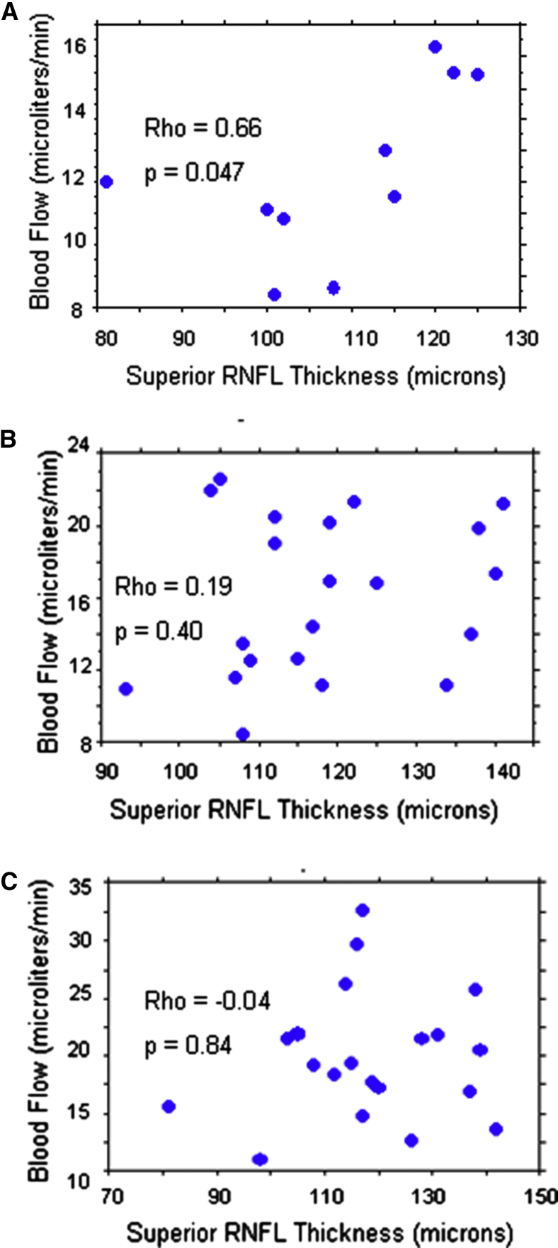
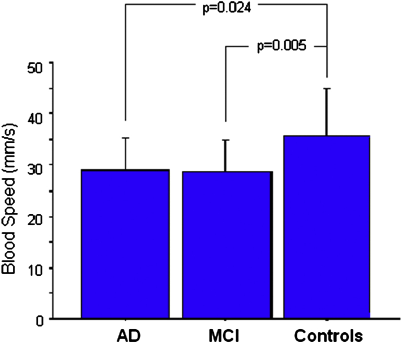
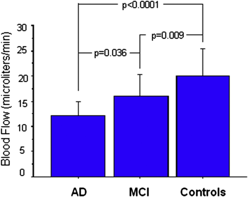
Blood column diameter in AD was narrower than in both MCI (P = .004) and controls (P = .002). However, blood speed in both AD (P = .024) and MCI (P = .005) was lower than in controls. As a result, the differences in blood flow between AD and MCI (P = .036), AD and controls (P < .0001), and MCI and controls (P = .009) were significant. Although there were no differences in RNFL thickness among the groups, blood flow was correlated (P = .047) with superior RNFL thickness in the AD group, but not in the MCI (P = .40) or control (P = .84) groups.
Retinal blood flow in MCI is intermediate between what is measured in control subjects and in AD patients. Our findings suggest that blood flow abnormalities may precede the neurodegeneration in AD.
与对照组相比,阿尔茨海默病(AD)患者表现出视网膜静脉变窄和视网膜静脉血流量减少。我们评估了这些异常是否存在于轻度认知障碍(MCI)患者中。
在确定全球临床痴呆评定量表后,52名受试者(10名AD患者、21名MCI患者和21名正常对照)接受了视网膜血流动力学分析。使用视网膜激光多普勒血流仪测量颞侧主要视网膜静脉的血柱直径、血流速度和血流量。此外,使用光学相干断层扫描测量视盘周围视网膜神经纤维层(RNFL)厚度。
AD患者的血柱直径比MCI患者(P = 0.004)和对照组(P = 0.002)更窄。然而,AD患者(P = 0.024)和MCI患者(P = 0.005)的血流速度均低于对照组。结果,AD与MCI(P = 0.036)、AD与对照组(P < 0.0001)以及MCI与对照组(P = 0.009)之间的血流量差异具有统计学意义。尽管各组之间RNFL厚度没有差异,但AD组的血流量与上方RNFL厚度相关(P = 0.047),而MCI组(P = 0.40)或对照组(P = 0.84)则无此相关性。
MCI患者的视网膜血流量介于对照组和AD患者之间。我们的研究结果表明,血流量异常可能先于AD中的神经退行性变出现。