Wang Meijuan, Zhu Yinbo, Shi Zhongyong, Li Chunbo, Shen Yuan
Department of Psychiatry, Tenth People's Hospital of Tong Ji University, Shanghai, China.
Shanghai Key Laboratory of Psychotic Disorders, Shanghai Mental Health Center, Shanghai Jiao Tong University School of Medicine, Shanghai, China.
Shanghai Arch Psychiatry. 2015 Oct;27(5):263-79. doi: 10.11919/j.issn.1002-0829.215100.
Previous studies report that the thickness of the peripheral retinal nerve fiber layer (RNFL) in individuals with Alzheimer's disease (AD) and mild cognitive impairment (MCI) is significantly thinner than in normal controls (NC), but RNFL thickness in different quadrants of the optic nerve remains unclear.
Conduct a systematic review of studies that assess peripheral RNFL thickness in AD and MCI.
Based on pre-defined criteria, studies in English or Chinese were identified from PubMed, Embase, ISI web of knowledge, Ovid/Medline, Science Direct, Cochrane Library, Chinese National Knowledge Infrastructure (CNKI), Chongqing VIP database, WANFANG DATA, and the China BioMedical Literature Service System (SinoMed). Review Manager 5.3 was used for analysis.
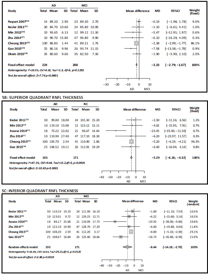
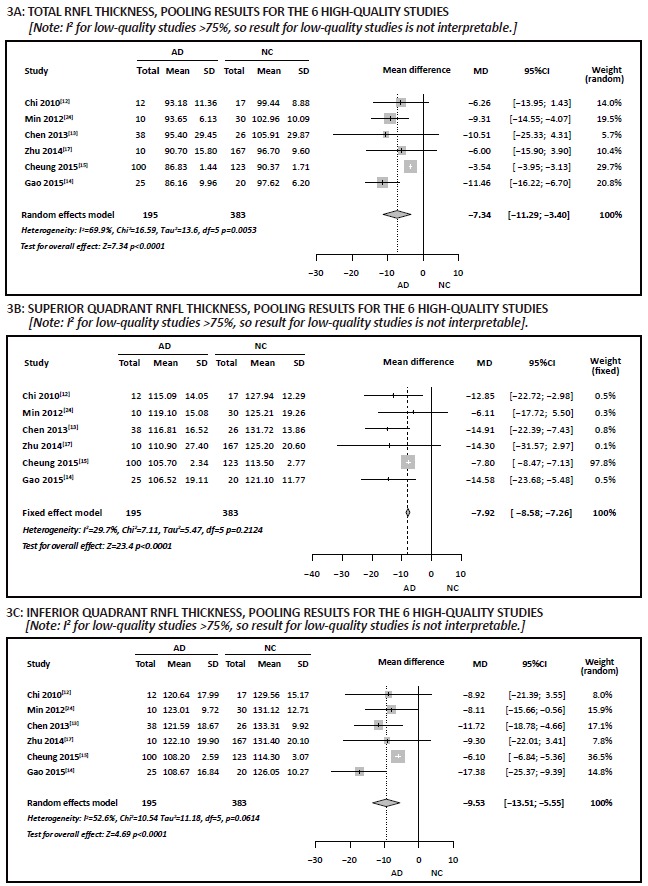
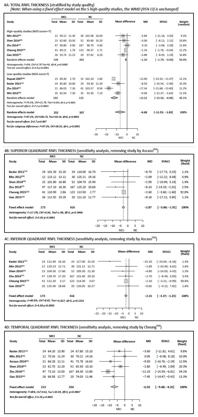
The 19 cross-sectional studies identified had a pooled sample of 1455 individuals. There was substantial heterogeneity between studies that compared RNFL in AD or MCI to normal controls, but this heterogeneity was primarily restricted to low-quality studies. Combining 6 high-quality studies (n=578) indicated that total RNFL thickness and the thickness of superior and inferior RNFL quadrants in AD were significantly thinner than in normal controls. Similarly, combining 5 high-quality studies (n=541) indicated significantly thinner total RNFL thickness in MCI than in controls. Six studies (n=589) found thinner RNFL in the superior and inferior quadrants in MCI than in controls;and 6 studies (n=487) found thinner RNFL in the temporal quadrant in MCI than in controls. Finally, 7 studies (n=432) indicated that total RNFL was thinner in AD than in MCI, and 6 studies (n=364) indicated thinner RNFL in the superior and inferior quadrants in AD than in MCI.
Much of the heterogeneity in results from previous studies may be due to poor methodology. Peripheral RNFL thicknesses, particularly in the superior and inferior quadrants, becomes progressively thinner as cognitive function declines, so this could be a candidate biomarker for early identification of AD. Methodologically rigorous studies in large population-based cohort studies that follow elderly individuals over time and that simultaneously collect information on potential mediating factors (such as blood pressure, blood glucose, and lipid levels) are needed to confirm or disprove the potential predictive value of RNFL.
以往研究报告称,阿尔茨海默病(AD)和轻度认知障碍(MCI)患者的周边视网膜神经纤维层(RNFL)厚度明显薄于正常对照(NC),但视神经不同象限的RNFL厚度仍不明确。
对评估AD和MCI患者周边RNFL厚度的研究进行系统评价。
根据预先设定的标准,从PubMed、Embase、ISI知识网络、Ovid/Medline、Science Direct、Cochrane图书馆、中国知网(CNKI)、重庆维普数据库、万方数据和中国生物医学文献服务系统(SinoMed)中检索英文或中文研究。使用Review Manager 5.3进行分析。
纳入的19项横断面研究共有1455名受试者。将AD或MCI患者的RNFL与正常对照进行比较的研究之间存在显著异质性,但这种异质性主要局限于低质量研究。合并6项高质量研究(n = 578)表明,AD患者的总RNFL厚度以及RNFL上下象限的厚度明显薄于正常对照。同样,合并5项高质量研究(n = 541)表明,MCI患者的总RNFL厚度明显薄于对照。6项研究(n = 589)发现,MCI患者RNFL上下象限的厚度薄于对照;6项研究(n = 487)发现,MCI患者RNFL颞侧象限的厚度薄于对照。最后,7项研究(n = 432)表明,AD患者的总RNFL薄于MCI患者,6项研究(n = 364)表明,AD患者RNFL上下象限的厚度薄于MCI患者。
以往研究结果的许多异质性可能归因于方法学不佳。周边RNFL厚度,尤其是上下象限的厚度,会随着认知功能下降而逐渐变薄,因此这可能是早期识别AD的候选生物标志物。需要在基于人群的大型队列研究中进行方法学严谨的研究,长期跟踪老年人,并同时收集潜在中介因素(如血压、血糖和血脂水平)的信息,以证实或反驳RNFL的潜在预测价值。