Ademola-Popoola Dupe S, Nzeh Donald A, Saka Sadiat E, Olokoba Lateefat B, Obajolowo Tokunbo S
Department of Radiology, University of Ilorin, Nigeria; Department of Ophthalmology, University of Ilorin Teaching Hospital, Nigeria.
Department of Radiology, University of Ilorin, Nigeria.
J Curr Ophthalmol. 2016 Feb 9;27(3-4):110-4. doi: 10.1016/j.joco.2015.12.002. eCollection 2015 Sep-Dec.
The study compared ocular biometry values using applanation and immersion techniques to determine the most applicable method for our tertiary training centre where personnel with different levels of experience and expertise in biometry take measurements used in calculation of required intraocular lens before cataract surgery.
The study was a prospective cross-sectional comparative study of different techniques of ocular biometry from diagnostic equipment (biometry probe 10 MHz Sonomed(®) A-scan (PACSCAN 300A, USA). Measurement variables were obtained among children and adults undergoing cataract surgery. Scleral (Prager) shell was used for the immersion technique followed by the contact technique by the same examiner.
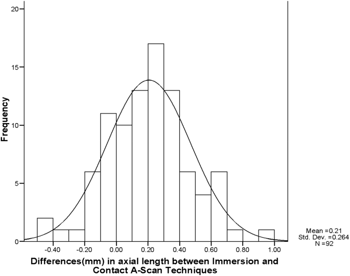
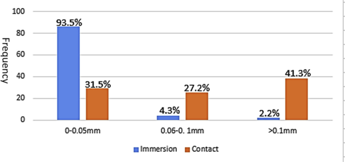
The biometry values of 92 eyes of 92 adult were taken. Their ages ranged from 18 to 95 years with a mean of 64.7 (SD ± 12.9) years. There were 55 (59.8%) males and 37 (40.2%) females, with a male to female ratio of 1.5:1. Average axial length (22.0-24.4 mm) eyes were the most common eyes measured in 75 (81.5%) of the cases. The means of the axial lengths biometry values with immersion and contact technique were 23.66(±1.36) and 23.46 mm (±1.46); the axial length differences was 0.2 ± 0.26 mm (range 0.0-0.94 mm) and statistically significant (95% CI of the Difference 0.15 to 0.26, p = 0.000). The Standard deviation SD (mm) of Individual Eye Axial Length showed a mean of 0.03 ± 0.04 (0.0-0.3) mm for immersion and for contact technique 0.14 ± 0.12(0.0-0.6)mm.
There was a significant difference in ocular biometry measurement with the contact and immersion ultrasound techniques. The immersion technique had better repeatability, thus it is ideal in a training hospital setting in a typical in sub-Saharan Africa who have limited resources to employ a dedicated person to do biometry; and where the different operators of A-scan machines have different levels of experience and expertise.
本研究比较了应用压平式和浸没式技术测量眼生物特征参数,以确定对于我们的三级培训中心而言哪种方法最为适用。在该培训中心,进行白内障手术所需人工晶状体计算时,由经验和专业水平各异的人员进行生物特征参数测量。
本研究是一项前瞻性横断面比较研究,采用诊断设备(10MHz Sonomed® A超生物测量探头(PACSCAN 300A,美国))的不同眼生物测量技术。测量变量取自接受白内障手术的儿童和成人。由同一名检查者先使用巩膜(普拉格)壳进行浸没式测量,随后采用接触式测量。
对92例成人的92只眼进行了生物特征参数测量。他们的年龄在18至95岁之间,平均年龄为64.7(标准差±12.9)岁。男性55例(59.8%),女性37例(40.2%),男女比例为1.5:1。轴长在22.0 - 24.4mm的眼睛是最常见的测量对象,共75例(81.5%)。浸没式和接触式技术测量的轴长生物特征参数均值分别为23.66(±1.36)和23.46mm(±1.46);轴长差异为0.2±0.26mm(范围0.0 - 0.94mm),具有统计学意义(差异的95%置信区间为0.15至0.26,p = 0.000)。单眼轴长的标准差(mm)显示,浸没式技术的均值为0.03±0.04(0.0 - 0.3)mm,接触式技术为0.14±0.12(0.0 - 0.6)mm。
接触式和浸没式超声技术在眼生物测量中存在显著差异。浸没式技术具有更好的重复性,因此对于撒哈拉以南非洲典型的资源有限、无法聘请专人进行生物测量的培训医院,以及A超机器操作人员经验和专业水平各异的情况而言,是理想的选择。