Shao Pei-Lin, Liao Jiunn-Der, Wong Tak-Wah, Wang Yi-Cheng, Leu Steve, Yip Hon-Kan
Department of Materials Science and Engineering, National Cheng Kung University, Tainan 70101, Taiwan.
Medical Device Innovation Center, National Cheng Kung University, Tainan 70101, Taiwan.
PLoS One. 2016 Jun 1;11(6):e0156699. doi: 10.1371/journal.pone.0156699. eCollection 2016.
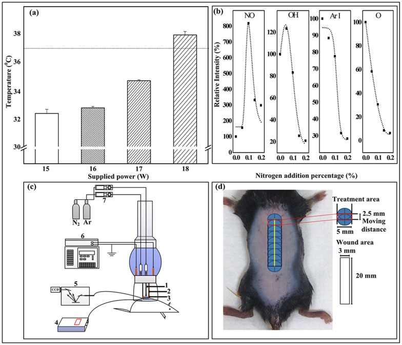
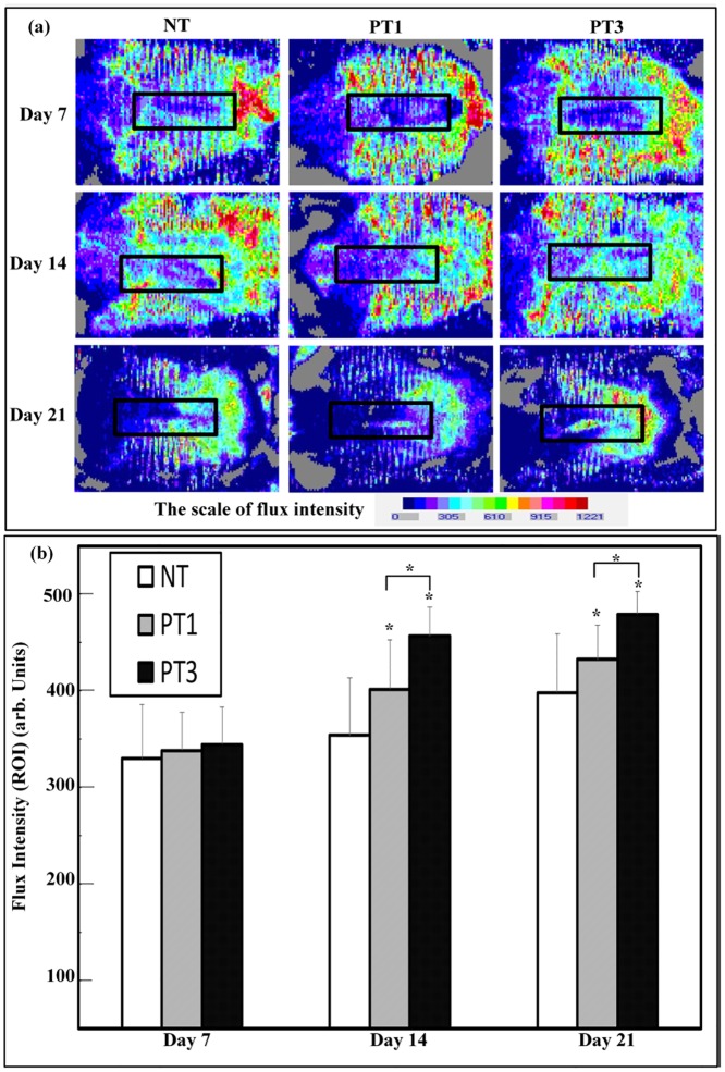
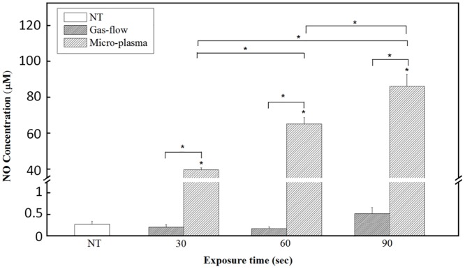
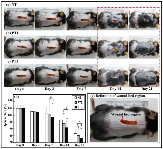
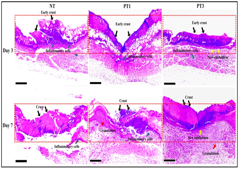
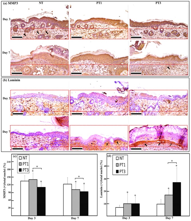
Micro-plasma is a possible alternative treatment for wound management. The effect of micro-plasma on wound healing depends on its composition and temperature. The authors previously developed a capillary-tube-based micro-plasma system that can generate micro-plasma with a high nitric oxide-containing species composition and mild working temperature. Here, the efficacy of micro-plasma treatment on wound healing in a laser-induced skin wound mouse model was investigated. A partial thickness wound was created in the back skin of each mouse and then treated with micro-plasma. Non-invasive methods, namely wound closure kinetics, optical coherence tomography (OCT), and laser Doppler scanning, were used to measure the healing efficiency in the wound area. Neo-tissue growth and the expressions of matrix metallopeptidase-3 (MMP-3) and laminin in the wound area were assessed using histological and immunohistochemistry (IHC) analysis. The results show that micro-plasma treatment promoted wound healing. Micro-plasma treatment significantly reduced the wound bed region. The OCT images and histological analysis indicates more pronounced tissue regrowth in the wound bed region after micro-plasma treatment. The laser Doppler images shows that micro-plasma treatment promoted blood flow in the wound bed region. The IHC results show that the level of laminin increased in the wound bed region after micro-plasma treatment, whereas the level of MMP-3 decreased. Based on these results, micro-plasma has potential to be used to promote the healing of skin wounds clinically.
微等离子体是伤口处理的一种可能的替代治疗方法。微等离子体对伤口愈合的影响取决于其成分和温度。作者之前开发了一种基于毛细管的微等离子体系统,该系统可以产生具有高含氮氧化物成分和温和工作温度的微等离子体。在此,研究了微等离子体处理对激光诱导皮肤伤口小鼠模型伤口愈合的疗效。在每只小鼠的背部皮肤制造一个部分厚度的伤口,然后用微等离子体进行处理。采用非侵入性方法,即伤口闭合动力学、光学相干断层扫描(OCT)和激光多普勒扫描,来测量伤口区域的愈合效率。使用组织学和免疫组织化学(IHC)分析评估伤口区域的新组织生长以及基质金属肽酶-3(MMP-3)和层粘连蛋白的表达。结果表明,微等离子体处理促进了伤口愈合。微等离子体处理显著减少了伤口床区域。OCT图像和组织学分析表明,微等离子体处理后伤口床区域的组织再生更为明显。激光多普勒图像显示,微等离子体处理促进了伤口床区域的血流。IHC结果显示,微等离子体处理后伤口床区域的层粘连蛋白水平升高,而MMP-3水平降低。基于这些结果,微等离子体在临床上有促进皮肤伤口愈合的潜力。