Boeckle Markus, Liegl Gregor, Jank Robert, Pieh Christoph
Department of Psychotherapy and Biopsychosocial Health, Danube University Krems, Dr.-Karl-Dorrek-Str. 30, 3500, Krems, Austria.
Department of Cognitive Biology, University of Vienna, Vienna, Austria.
BMC Psychiatry. 2016 Jun 10;16:195. doi: 10.1186/s12888-016-0890-x.
Conversion Disorders (CD) are prevalent functional disorders. Although the pathogenesis is still not completely understood, an interaction of genetic, neurobiological, and psychosocial factors is quite likely. The aim of this study is to provide a systematic overview on imaging studies on CDs and investigate neuronal areas involved in Motor Conversion Disorders (MCD).
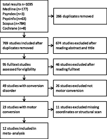
A systematic literature search was conducted on CD. Subsequently a meta-analysis of functional neuroimaging studies on MCD was implemented using an Activation Likelihood Estimation (ALE). We calculated differences between patients and healthy controls as well as between affected versus unaffected sides in addition to an overall analysis in order to identify neuronal areas related to MCD.
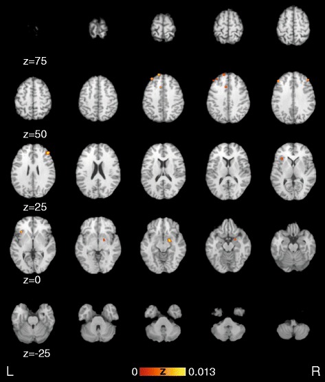
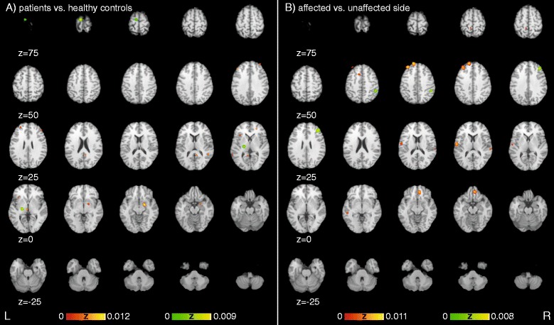
Patients with MCD differ from healthy controls in the amygdala, superior temporal lobe, retrosplenial area, primary motor cortex, insula, red nucleus, thalamus, anterior as well as dorsolateral prefrontal and frontal cortex. When comparing affected versus unaffected sides, temporal cortex, dorsal anterior cingulate cortex, supramarginal gyrus, dorsal temporal lobe, anterior insula, primary somatosensory cortex, superior frontal gyrus and anterior prefrontal as well as frontal cortex show significant differences.
Neuronal areas seem to be involved in the pathogenesis, maintenance or as a result of MCD. Areas that are important for motor-planning, motor-selection or autonomic response seem to be especially relevant. Our results support the emotional unawareness theory but also underline the need of more support by conduction imaging studies on both CD and MCD.
转换障碍(CD)是常见的功能性障碍。尽管其发病机制仍未完全明确,但遗传、神经生物学和心理社会因素之间的相互作用很可能存在。本研究的目的是对转换障碍的影像学研究进行系统综述,并调查与运动性转换障碍(MCD)相关的神经区域。
对转换障碍进行了系统的文献检索。随后,使用激活可能性估计(ALE)对运动性转换障碍的功能性神经影像学研究进行了荟萃分析。除了进行整体分析以识别与运动性转换障碍相关的神经区域外,我们还计算了患者与健康对照之间以及患侧与未患侧之间的差异。
运动性转换障碍患者在杏仁核、颞上叶、压后皮质、初级运动皮层、脑岛、红核、丘脑、前额叶和额叶背外侧前部与健康对照存在差异。在比较患侧与未患侧时,颞叶皮质、背侧前扣带回皮质、缘上回、颞叶背侧、脑岛前部、初级躯体感觉皮层、额上回、前额叶前部以及额叶皮质显示出显著差异。
神经区域似乎参与了运动性转换障碍的发病机制、维持过程或其结果。对运动计划、运动选择或自主反应重要的区域似乎尤其相关。我们的结果支持情绪无意识理论,但也强调了需要通过对转换障碍和运动性转换障碍的传导影像学研究提供更多支持。