Department of Neurology, University of Texas McGovern Medical School at Houston, Houston, TX, 77030, USA.
Department of Anesthesiology and Center for Shock, Trauma and Anesthesiology Research (STAR), University of Maryland School of Medicine, Baltimore, MD, 21201, USA.
J Neuroinflammation. 2018 May 17;15(1):148. doi: 10.1186/s12974-018-1188-3.
Activation of transforming growth factor-β-activated kinase 1 (TAK1) occurs after stroke and leads to an exacerbation of brain injury. TAK1 is involved in innate and adaptive immune responses, but it has divergent inflammatory effects that are dependent on the cell type in which it is activated. There is a robust infiltration of myeloid cells after stroke; however, the contribution of myeloid TAK1 to cerebral ischemia is currently unknown. We hypothesized that myeloid-specific deletion of TAK1 would protect against ischemic brain injury.
Myeloid TAK1M and wild-type (WT) mice were subjected to middle cerebral artery occlusion (MCAo). Brain-infiltrating and splenic immune cells were evaluated at 3 days after stroke. Assessment of infarct size and behavioral deficits were performed on days 3 and 7 post-stroke.
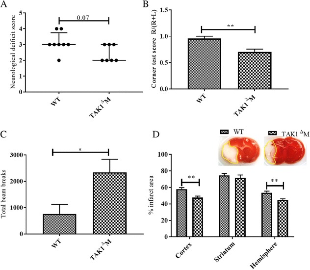
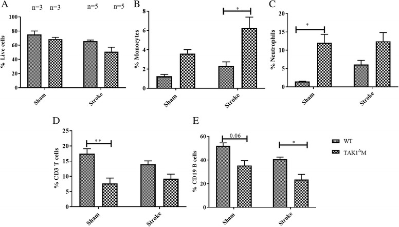
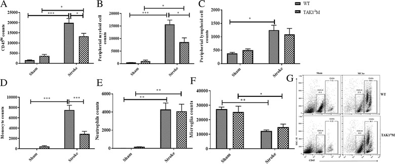
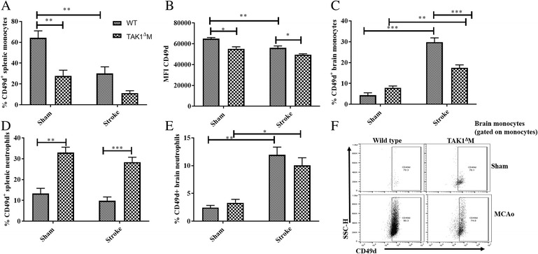
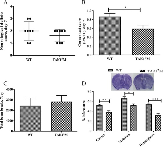
Infarcts were significantly smaller in TAK1M mice (p < 0.01), and behavioral deficits were less severe despite equivalent reduction in cerebral blood flow. Flow cytometry demonstrated an increase in the frequency of splenic monocytes and neutrophils (p < 0.05) and a decrease in splenic CD3 T (p < 0.01) and CD19 B (p = 0.06) cells in TAK1M mice compared to WT at baseline. Three days after stroke, a significant increase in the number of brain-infiltrating immune cell was observed in both TAK1M (p < 0.05) and WT (p < 0.001) mice compared to their respective shams. However, there was a significant decrease in the infiltrating CD45 immune cell counts (p < 0.05), with a pronounced reduction in infiltrating monocytes (p < 0.001) in TAK1M after stroke compared to WT stroke mice. Additionally, a significant reduction in CD49d monocytes was seen in the brains of TAK1M stroke mice compared to wild-type mice. Importantly, TAK1M MCAo mice had smaller infarcts and improved behavioral outcomes at day 7 post-stroke.
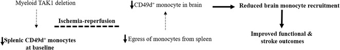
Our results showed that deletion of myeloid TAK1 resulted in smaller infarcts and improved functional outcomes at the peak of inflammation (day 3) and a reduction in brain-infiltrating immune cells that were primarily monocytes. Myeloid TAK1 deletion was also protective at 7 days post MCAo, reflecting a detrimental role of myeloid TAK1 in the progression of ischemic injury.
转化生长因子-β激活激酶 1(TAK1)在中风后被激活,导致脑损伤加重。TAK1 参与先天和适应性免疫反应,但它具有依赖于被激活的细胞类型的不同炎症效应。中风后会有大量髓样细胞浸润;然而,髓样 TAK1 对脑缺血的贡献目前尚不清楚。我们假设髓样细胞特异性敲除 TAK1 将保护对抗缺血性脑损伤。
TAK1M 和野生型(WT)小鼠接受大脑中动脉闭塞(MCAo)。在中风后 3 天评估脑内浸润和脾免疫细胞。在中风后第 3 天和第 7 天进行梗死面积和行为缺陷评估。
TAK1M 小鼠的梗死灶明显较小(p<0.01),尽管脑血流减少程度相同,但行为缺陷较轻。流式细胞术显示,与 WT 相比,TAK1M 小鼠的脾单核细胞和中性粒细胞频率增加(p<0.05),脾 CD3 T(p<0.01)和 CD19 B(p=0.06)细胞减少。与 WT 相比,TAK1M 小鼠在基线时脑内浸润免疫细胞数量显著增加(p<0.05)。中风后 3 天,与各自的假手术相比,TAK1M(p<0.05)和 WT (p<0.001)小鼠的脑内浸润免疫细胞数量均显著增加。然而,浸润性 CD45 免疫细胞计数显著减少(p<0.05),中风后 TAK1M 小鼠浸润性单核细胞明显减少(p<0.001)。此外,与野生型小鼠相比,TAK1M 中风小鼠大脑中的 CD49d 单核细胞显著减少。重要的是,TAK1M MCAo 小鼠在中风后第 7 天的梗死灶较小,行为学结果改善。
我们的结果表明,髓样细胞 TAK1 的缺失导致中风后炎症高峰期(第 3 天)梗死灶较小,以及浸润性免疫细胞减少,主要是单核细胞。TAK1M 缺失在 MCAo 后 7 天也具有保护作用,反映了髓样 TAK1 在缺血性损伤进展中的有害作用。