Zheng Yong-Ping, Lee Timothy Tin-Yan, Lai Kelly Ka-Lee, Yip Benjamin Hon-Kei, Zhou Guang-Quan, Jiang Wei-Wei, Cheung James Chung-Wai, Wong Man-Sang, Ng Bobby King-Wah, Cheng Jack Chun-Yiu, Lam Tsz-Ping
Interdisciplinary Division of Biomedical Engineering, The Hong Kong Polytechnic University, Hong Kong, People's Republic of China.
School of Public Health and Primary Care, The Chinese University of Hong Kong, Hong Kong, People's Republic of China.
Scoliosis Spinal Disord. 2016 May 31;11:13. doi: 10.1186/s13013-016-0074-y. eCollection 2016.
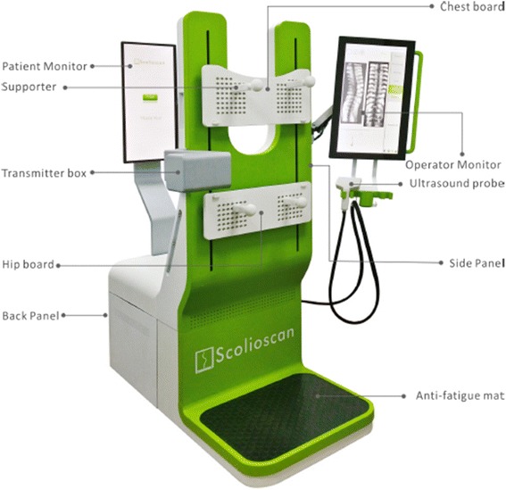
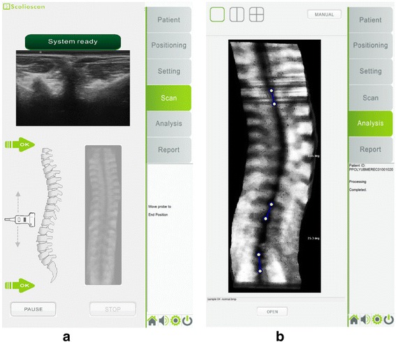
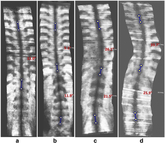
Radiographic evaluation for patients with scoliosis using Cobb method is the current gold standard, but radiography has radiation hazards. Several groups have recently demonstrated the feasibility of using 3D ultrasound for the evaluation of scoliosis. Ultrasound imaging is radiation-free, comparatively more accessible, and inexpensive. However, a reliable and valid 3D ultrasound system ready for clinical scoliosis assessment has not yet been reported. Scolioscan is a newly developed system targeted for scoliosis assessment in clinics by using coronal images of spine generated by a 3D ultrasound volume projection imaging method. The aim of this study is to test the reliability of spine deformity measurement of Scolioscan and its validity compared to the gold standard Cobb angle measurements from radiography in adolescent idiopathic scoliosis (AIS) patients.
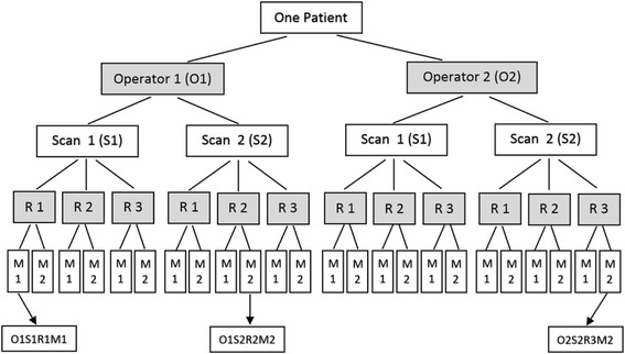
Prospective study divided into two stages: 1) Investigation of intra- and inter- reliability between two operators for acquiring images using Scolioscan and among three raters for measuring spinal curves from those images; 2) Correlation between the Cobb angle obtained from radiography by a medical doctor and the spine curve angle obtained using Scolioscan (Scolioscan angle). The raters for ultrasound images and the doctors for evaluating radiographic images were mutually blinded. The two stages of tests involved 20 (80 % females, total of 26 angles, age of 16.4 ± 2.7 years, and Cobb angle of 27.6 ± 11.8°) and 49 (69 % female, 73 angles, 15.8 ± 2.7 years and 24.8 ± 9.7°) AIS patients, respectively. Intra-class correlation coefficients (ICC) and Bland-Altman plots and root-mean-square differences (RMS) were employed to determine correlations, which interpreted based on defined criteria.
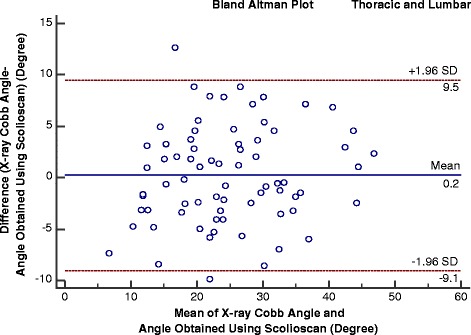
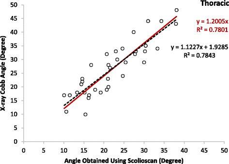
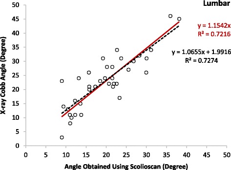
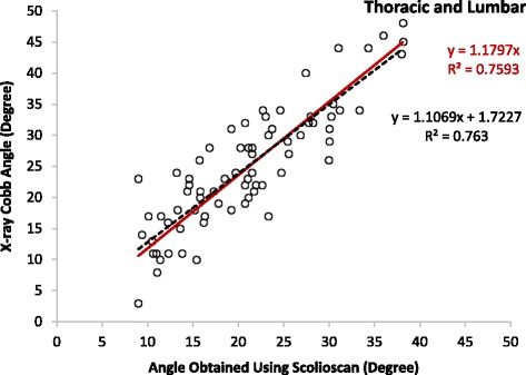
We demonstrated a very good intra-rater and intra-operator reliability for Scolioscan angle measurement with ICC larger than 0.94 and 0.88, respectively. Very good inter-rater and inter-operator reliability was also demonstrated, with both ICC larger than 0.87. For the thoracic deformity measurement, the RMS were 2.5 and 3.3° in the intra- and inter-operator tests, and 1.5 and 3.6° in the intra- and inter-rater tests, respectively. The RMS differences were 3.1, 3.1, 1.6, 3.7° in the intra- and inter-operator and intra- and inter-rater tests, respectively, for the lumbar angle measurement. Moderate to strong correlations (R(2) > 0.72) were observed between the Scolioscan angles and Cobb angles for both the thoracic and lumbar regions. It was noted that the Scolioscan angle slightly underestimated the spinal deformity in comparison with Cobb angle, and an overall regression equation y = 1.1797x (R(2) = 0.76) could be used to translate the Scolioscan angle (x) to Cobb angle (y) for this group of patients. The RMS difference between Scolioscan angle and Cobb angle was 4.7 and 6.2°, with and without the correlation using the overall regression equation.
We showed that Scolioscan is reliable for measuring coronal deformity for patients with AIS and appears promising in screening large numbers of patients, for progress monitoring, and evaluation of treatment outcomes. Due to it being radiation-free and relatively low-cost, Scolioscan has potential to be widely implemented and may contribute to reducing radiation dose during serial monitoring.
使用Cobb法对脊柱侧弯患者进行影像学评估是当前的金标准,但放射检查存在辐射危害。最近有几个研究小组证明了使用三维超声评估脊柱侧弯的可行性。超声成像无辐射,相对更易获得且成本低廉。然而,尚未有可靠且有效的三维超声系统可用于临床脊柱侧弯评估的报道。Scolioscan是一种新开发的系统,旨在通过三维超声容积投影成像方法生成的脊柱冠状面图像,对临床中的脊柱侧弯进行评估。本研究的目的是测试Scolioscan测量脊柱畸形的可靠性,以及与青少年特发性脊柱侧弯(AIS)患者放射检查的金标准Cobb角测量结果相比的有效性。
前瞻性研究分为两个阶段:1)两名操作人员使用Scolioscan获取图像之间以及三名评估者测量这些图像脊柱曲线之间的组内和组间可靠性研究;2)医生通过放射检查获得的Cobb角与使用Scolioscan获得的脊柱曲线角度(Scolioscan角)之间的相关性研究。超声图像评估者和放射图像评估医生相互不知情。两个测试阶段分别纳入了20例(80%为女性,共26个角度,年龄16.4±2.7岁,Cobb角27.6±11.8°)和49例(69%为女性,73个角度,15.8±2.7岁,24.8±9.7°)AIS患者。采用组内相关系数(ICC)、Bland-Altman图和均方根差(RMS)来确定相关性,并根据既定标准进行解读。
我们证明了Scolioscan角度测量具有非常好的评估者内和操作人员内可靠性,ICC分别大于0.94和0.88。也证明了评估者间和操作人员间具有非常好的可靠性,ICC均大于0.87。对于胸椎畸形测量,操作人员内和操作人员间测试的RMS分别为2.5°和3.3°,评估者内和评估者间测试的RMS分别为1.5°和3.6°。对于腰椎角度测量,操作人员内和操作人员间以及评估者内和评估者间测试的RMS差异分别为3.1°、3.1°、1.6°、3.7°。胸椎和腰椎区域的Scolioscan角与Cobb角之间均观察到中度至高度相关性(R²>0.72)。值得注意的是,与Cobb角相比,Scolioscan角略微低估了脊柱畸形,对于该组患者,可使用总体回归方程y = 1.1797x(R² = 0.76)将Scolioscan角(x)转换为Cobb角(y)。使用总体回归方程和不使用总体回归方程时,Scolioscan角与Cobb角之间的RMS差异分别为4.7°和6.2°。
我们表明Scolioscan对于测量AIS患者冠状面畸形是可靠的,并且在筛查大量患者、进展监测和治疗效果评估方面似乎很有前景。由于其无辐射且成本相对较低,Scolioscan有潜力被广泛应用,并可能有助于在连续监测期间减少辐射剂量。