Zhang Fan, Suh Kyung-Jin, Lee Kyung-Min
Department of Orthodontics, School of Dentistry, 4D Dental Research Institute, Chonnam National University, Gwangju, Korea.
PLoS One. 2016 Jun 15;11(6):e0157713. doi: 10.1371/journal.pone.0157713. eCollection 2016.
Dental measurements have been commonly taken from plaster dental models obtained from alginate impressions can. Through the use of an intraoral scanner, digital impressions now acquire the information directly from the mouth. The purpose of this study was to determine the validity of the intraoral scans compared to plaster models.
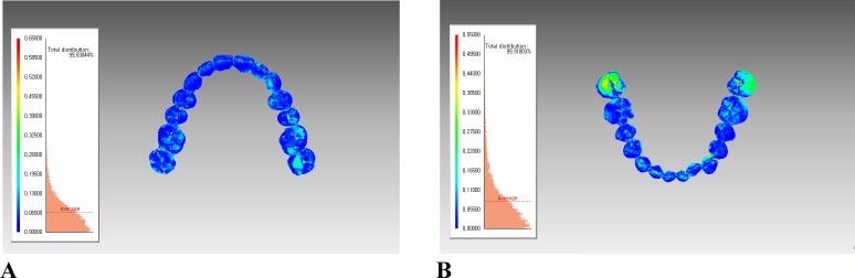
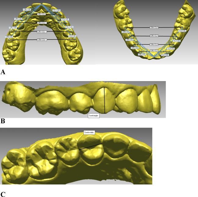
Two types of dental models (intraoral scan and plaster model) of 20 subjects were included in this study. The subjects had impressions taken of their teeth and made as plaster model. In addition, their mouths were scanned with the intraoral scanner and the scans were converted into digital models. Eight transverse and 16 anteroposterior measurements, 24 tooth heights and widths were recorded on the plaster models with a digital caliper and on the intraoral scan with 3D reverse engineering software. For 3D surface analysis, the two models were superimposed by using best-fit algorithm. The average differences between the two models at all points on the surfaces were computed. Paired t-test and Bland-Altman plot were used to determine the validity of measurements from the intraoral scan compared to those from the plaster model.
There were no significant differences between the plaster models and intraoral scans, except for one measurement of lower intermolar width. The Bland-Altman plots of all measurements showed that differences between the two models were within the limits of agreement. The average surface difference between the two models was within 0.10 mm.
The results of the present study indicate that the intraoral scans are clinically acceptable for diagnosis and treatment planning in dentistry and can be used in place of plaster models.
牙科测量通常是从藻酸盐印模制取的石膏牙模型上进行的。通过使用口腔内扫描仪,数字印模现在可以直接从口腔获取信息。本研究的目的是确定与石膏模型相比口腔内扫描的有效性。
本研究纳入了20名受试者的两种类型的牙模型(口腔内扫描模型和石膏模型)。对受试者的牙齿制取印模并制作成石膏模型。此外,用口腔内扫描仪对他们的口腔进行扫描,并将扫描结果转换为数字模型。使用数字卡尺在石膏模型上记录8项横向和16项前后向测量值、24项牙齿高度和宽度,并用三维逆向工程软件在口腔内扫描模型上记录这些数据。对于三维表面分析,使用最佳拟合算法将两种模型叠加。计算两种模型在表面所有点的平均差异。采用配对t检验和布兰德-奥特曼图来确定与石膏模型相比口腔内扫描测量的有效性。
除一项下颌磨牙间宽度测量外,石膏模型和口腔内扫描之间没有显著差异。所有测量的布兰德-奥特曼图显示,两种模型之间的差异在一致限度内。两种模型的平均表面差异在0.10毫米以内。
本研究结果表明,口腔内扫描在牙科诊断和治疗计划中在临床上是可接受的,并且可以替代石膏模型使用。