Ibrahim Mohd Rafiq Mohd, Singh Simmrat, Merican Azhar Mahmood, Raghavendran Hanumantha Rao Balaji, Murali Malliga Raman, Naveen Sangeetha Vasudevaraj, Kamarul Tunku
Tissue Engineering Group (TEG), National Orthopaedic Centre of Excellence in Research and Learning (NOCERAL), Department of Orthopaedic Surgery, Faculty of Medicine, University of Malaya, Kuala Lumpur, Malaysia.
Clinical Investigative Centre, Faculty of Medicine, University Malaya Medical Center, Kuala Lumpur, Malaysia.
BMC Vet Res. 2016 Jun 16;12(1):112. doi: 10.1186/s12917-016-0724-6.
Fracture healing in bone gap is one of the major challenges encountered in Orthopedic Surgery. At present, the treatment includes bone graft, employing either internal or external fixation which has a significant impact on the patient, family and even society. New drugs are emerging in the markets such as anabolic bone-forming agents including teriparatide and strontium ranelate to stimulate bone growth. Based on the mechanism of their actions, we embarked on a study on the healing of a fractured ulna with bone gap in a rabbit model. We segregated ten rabbits into two groups: five rabbits in the test group and five rabbits in the control group. We created a 5 mm bone gap in the ulna bone, removing the periosteum as well. Rabbits in the test group received 450 mg/kg of strontium ranelate via oral administration, daily, for six weeks. The x-rays, CT scans and blood tests were performed every two weeks. At the end of six weeks, the rabbits were sacrificed, and the radius and ulna bones harvested for histopathological examination.
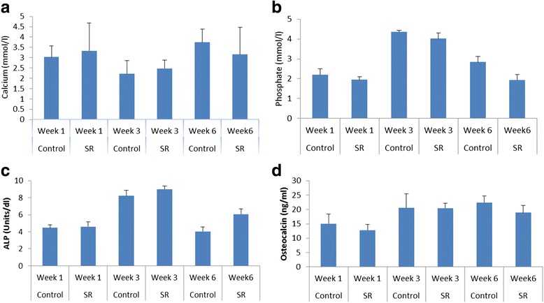
Based on the x-rays and CT scans, fracture healing or bone formation was observed to be faster in the control group. From the x-ray findings, 80 % of the fracture united and by CT scan, 60 % of the fracture united in the control group at the end of the six-week study. None of the fractures united in the test group. However, the histopathology report showed that a callus of different stages was being formed in both groups, consisting of 80 % of bone. The serum levels of osteocalcin and alkaline phosphatase initially remained similar up to three weeks and changed slightly at the end of six weeks.
We conclude that the strontium effect begins slowly, and while it may not interfere with bone cell proliferation it may interfere in the mineralization and delay the acute stage of fracture healing. We recommend that a larger sample size and a longer duration of the study period be implemented to confirm our finding.
骨间隙骨折愈合是骨科手术中面临的主要挑战之一。目前,治疗方法包括骨移植,采用内固定或外固定,这对患者、家庭乃至社会都有重大影响。市场上出现了一些新药,如促合成骨形成剂,包括特立帕肽和雷奈酸锶,以刺激骨生长。基于它们的作用机制,我们在兔模型上开展了一项关于尺骨骨间隙骨折愈合的研究。我们将10只兔子分为两组:实验组5只兔子,对照组5只兔子。我们在尺骨上制造了一个5毫米的骨间隙,并去除了骨膜。实验组的兔子每天口服450毫克/千克雷奈酸锶,持续六周。每两周进行一次X光、CT扫描和血液检查。六周结束时,处死兔子,取出桡骨和尺骨进行组织病理学检查。
根据X光和CT扫描,观察到对照组的骨折愈合或骨形成更快。从X光检查结果来看,在为期六周的研究结束时,对照组80%的骨折实现了愈合,通过CT扫描,60%的骨折实现了愈合。实验组没有骨折愈合。然而,组织病理学报告显示,两组均形成了不同阶段的骨痂,其中80%为骨组织。骨钙素和碱性磷酸酶的血清水平在最初三周内基本保持相似,六周结束时略有变化。
我们得出结论,锶的作用起效缓慢,虽然它可能不干扰骨细胞增殖,但可能干扰矿化并延迟骨折愈合的急性期。我们建议采用更大的样本量和更长的研究周期来证实我们的发现。