Gao Wei, Chen Shu-Rui, Wu Meng-Yao, Gao Kai, Li Yuan-Long, Wang Hong-Yu, Li Chen-Yuan, Li Hong
Department of Biochemistry, Liaoning Medical University, Jinzhou, Liaoning Province, China.
Neural Regen Res. 2016 May;11(5):823-8. doi: 10.4103/1673-5374.182711.
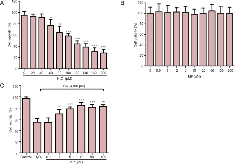
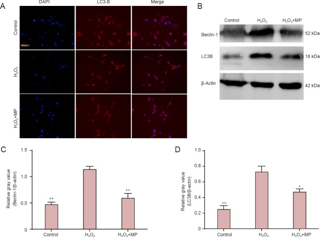
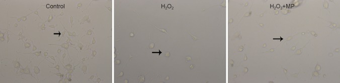
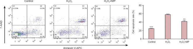
Methylprednisolone markedly reduces autophagy and apoptosis after secondary spinal cord injury. Here, we investigated whether pretreatment of cells with methylprednisolone would protect neuron-like cells from subsequent oxidative damage via suppression of autophagy and apoptosis. Cultured N2a cells were pretreated with 10 µM methylprednisolone for 30 minutes, then exposed to 100 µM H2O2 for 24 hours. Inverted phase contrast microscope images, MTT assay, flow cytometry and western blot results showed that, compared to cells exposed to 100 µM H2O2 alone, cells pretreated with methylprednisolone had a significantly lower percentage of apoptotic cells, maintained a healthy morphology, and showed downregulation of autophagic protein light chain 3B and Beclin-1 protein expression. These findings indicate that methylprednisolone exerted neuroprotective effects against oxidative damage by suppressing autophagy and apoptosis.
甲基强的松龙显著减少继发性脊髓损伤后的自噬和凋亡。在此,我们研究了用甲基强的松龙预处理细胞是否会通过抑制自噬和凋亡来保护神经元样细胞免受随后的氧化损伤。将培养的N2a细胞用10 µM甲基强的松龙预处理30分钟,然后暴露于100 µM过氧化氢中24小时。倒置相差显微镜图像、MTT法、流式细胞术和蛋白质印迹结果表明,与仅暴露于100 µM过氧化氢的细胞相比,用甲基强的松龙预处理的细胞凋亡细胞百分比显著降低,保持健康形态,并显示自噬蛋白轻链3B和Beclin-1蛋白表达下调。这些发现表明,甲基强的松龙通过抑制自噬和凋亡对氧化损伤发挥神经保护作用。