• 文献检索
  • 文档翻译
  • 深度研究
  • 学术资讯
  • Suppr Zotero 插件Zotero 插件
  • 邀请有礼
  • 套餐&价格
  • 历史记录
应用&插件
Suppr Zotero 插件Zotero 插件浏览器插件Mac 客户端Windows 客户端微信小程序
定价
高级版会员购买积分包购买API积分包
服务
文献检索文档翻译深度研究API 文档MCP 服务
关于我们
关于 Suppr公司介绍联系我们用户协议隐私条款
关注我们

Suppr 超能文献

核心技术专利:CN118964589B侵权必究
粤ICP备2023148730 号-1Suppr @ 2026

文献检索

告别复杂PubMed语法,用中文像聊天一样搜索,搜遍4000万医学文献。AI智能推荐,让科研检索更轻松。

立即免费搜索

文件翻译

保留排版,准确专业,支持PDF/Word/PPT等文件格式,支持 12+语言互译。

免费翻译文档

深度研究

AI帮你快速写综述,25分钟生成高质量综述,智能提取关键信息,辅助科研写作。

立即免费体验

惊恐障碍患者杏仁核的亚区域形态改变

Subregional Shape Alterations in the Amygdala in Patients with Panic Disorder.

作者信息

Yoon Sujung, Kim Jieun E, Kim Geon Ha, Kang Hee Jin, Kim Bori R, Jeon Saerom, Im Jooyeon Jamie, Hyun Heejung, Moon Sohyeon, Lim Soo Mee, Lyoo In Kyoon

机构信息

Ewha Brain Institute, Ewha Womans University, Seoul, South Korea.

Department of Brain and Cognitive Sciences, Ewha Womans University, Seoul, South Korea.

出版信息

PLoS One. 2016 Jun 23;11(6):e0157856. doi: 10.1371/journal.pone.0157856. eCollection 2016.

DOI:10.1371/journal.pone.0157856
PMID:27336300
原文链接:https://pmc.ncbi.nlm.nih.gov/articles/PMC4919046/
Abstract

BACKGROUND

The amygdala has been known to play a pivotal role in mediating fear-related responses including panic attacks. Given the functionally distinct role of the amygdalar subregions, morphometric measurements of the amygdala may point to the pathophysiological mechanisms underlying panic disorder. The current study aimed to determine the global and local morphometric alterations of the amygdala related to panic disorder.

METHODS

Volumetric and surface-based morphometric approach to high-resolution three-dimensional T1-weighted images was used to examine the structural variations of the amygdala, with respect to extent and location, in 23 patients with panic disorder and 31 matched healthy individuals.

RESULTS

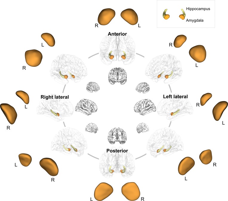
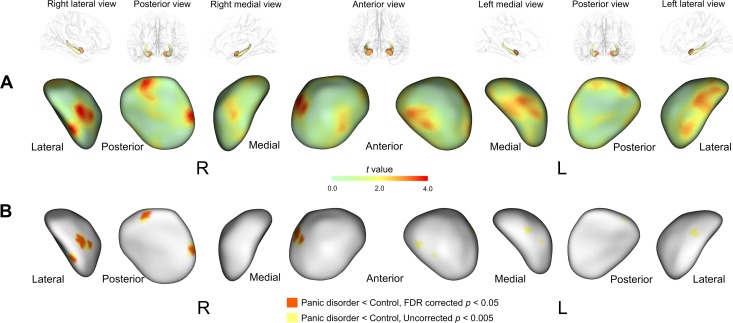
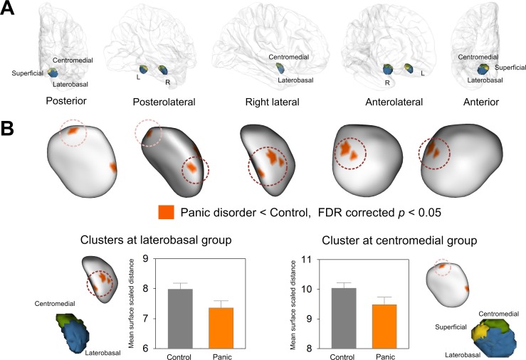
There were no significant differences in bilateral amygdalar volumes between patients with panic disorder and healthy individuals despite a trend-level right amygdalar volume reduction related to panic disorder (right, β = -0.23, p = 0.09, Cohen's d = 0.51; left, β = -0.18, p = 0.19, Cohen's d = 0.45). Amygdalar subregions were localized into three groups including the superficial, centromedial, and laterobasal groups based on the cytoarchitectonically defined probability map. Surface-based morphometric analysis revealed shape alterations in the laterobasal and centromedial groups of the right amygdala in patients with panic disorder (false discovery rate corrected p < 0.05).

CONCLUSIONS

The current findings suggest that subregion-specific shape alterations in the right amygdala may be involved in the development and maintenance of panic disorder, which may be attributed to the cause or effects of amygdalar hyperactivation.

摘要

背景

已知杏仁核在介导包括惊恐发作在内的与恐惧相关的反应中起关键作用。鉴于杏仁核亚区域功能不同,对杏仁核的形态测量可能指向惊恐障碍潜在的病理生理机制。本研究旨在确定与惊恐障碍相关的杏仁核整体和局部形态学改变。

方法

采用基于容积和表面的形态测量方法,对高分辨率三维T1加权图像进行分析,以检查23例惊恐障碍患者和31名匹配的健康个体中杏仁核在范围和位置方面的结构变化。

结果

惊恐障碍患者和健康个体之间双侧杏仁核体积无显著差异,尽管存在与惊恐障碍相关的右侧杏仁核体积呈趋势性减小(右侧,β = -0.23,p = 0.09,Cohen's d = 0.51;左侧,β = -0.18,p = 0.19,Cohen's d = 0.45)。根据细胞构筑学定义的概率图,杏仁核亚区域分为三组,包括浅表组、中央内侧组和外侧基底组。基于表面的形态测量分析显示,惊恐障碍患者右侧杏仁核的外侧基底组和中央内侧组存在形状改变(错误发现率校正p < 0.05)。

结论

目前的研究结果表明,右侧杏仁核特定亚区域的形状改变可能参与惊恐障碍的发生和维持,这可能归因于杏仁核过度激活的原因或结果。

https://cdn.ncbi.nlm.nih.gov/pmc/blobs/971f/4919046/2cfcd35b75c7/pone.0157856.g003.jpg
https://cdn.ncbi.nlm.nih.gov/pmc/blobs/971f/4919046/994edbca054d/pone.0157856.g001.jpg
https://cdn.ncbi.nlm.nih.gov/pmc/blobs/971f/4919046/166bb1beee49/pone.0157856.g002.jpg
https://cdn.ncbi.nlm.nih.gov/pmc/blobs/971f/4919046/2cfcd35b75c7/pone.0157856.g003.jpg
https://cdn.ncbi.nlm.nih.gov/pmc/blobs/971f/4919046/994edbca054d/pone.0157856.g001.jpg
https://cdn.ncbi.nlm.nih.gov/pmc/blobs/971f/4919046/166bb1beee49/pone.0157856.g002.jpg
https://cdn.ncbi.nlm.nih.gov/pmc/blobs/971f/4919046/2cfcd35b75c7/pone.0157856.g003.jpg

相似文献

1
Subregional Shape Alterations in the Amygdala in Patients with Panic Disorder.惊恐障碍患者杏仁核的亚区域形态改变
PLoS One. 2016 Jun 23;11(6):e0157856. doi: 10.1371/journal.pone.0157856. eCollection 2016.
2
Laterobasal amygdalar enlargement in 6- to 7-year-old children with autism spectrum disorder.6至7岁自闭症谱系障碍儿童的杏仁核后基底部增大
Arch Gen Psychiatry. 2010 Nov;67(11):1187-97. doi: 10.1001/archgenpsychiatry.2010.148.
3
Amygdalar shape analysis method using surface contour aligning, spherical mapping, and probabilistic subregional segmentation.使用表面轮廓配准、球面映射和概率亚区分割的杏仁核形状分析方法。
Neurosci Lett. 2011 Jan 13;488(1):65-9. doi: 10.1016/j.neulet.2010.11.005. Epub 2010 Nov 5.
4
Smaller amygdala is associated with anxiety in patients with panic disorder.杏仁核较小与惊恐障碍患者的焦虑有关。
Psychiatry Clin Neurosci. 2009 Jun;63(3):266-76. doi: 10.1111/j.1440-1819.2009.01960.x.
5
fMRI amygdala activation during a spontaneous panic attack in a patient with panic disorder.惊恐障碍患者自发惊恐发作期间的功能磁共振成像杏仁核激活
World J Biol Psychiatry. 2007;8(4):269-72. doi: 10.1080/15622970701216673.
6
Amygdalar atrophy in panic disorder patients detected by volumetric magnetic resonance imaging.通过容积磁共振成像检测惊恐障碍患者的杏仁核萎缩。
Neuroimage. 2003 May;19(1):80-90. doi: 10.1016/s1053-8119(03)00036-3.
7
Temporal and right frontal lobe alterations in panic disorder: a quantitative volumetric and voxel-based morphometric MRI study.惊恐障碍患者的时间和右额叶改变:一项定量容积和基于体素的形态测量磁共振成像研究。
Psychol Med. 2010 Nov;40(11):1879-86. doi: 10.1017/S0033291709991930. Epub 2010 Jan 8.
8
Smaller volumes in the lateral and basal nuclei of the amygdala in patients with panic disorder.惊恐障碍患者杏仁核外侧核和基底核的体积较小。
PLoS One. 2018 Nov 7;13(11):e0207163. doi: 10.1371/journal.pone.0207163. eCollection 2018.
9
Amygdalar hyperactivity, a fear-related link between panic disorder and mesiotemporal epilepsy.杏仁核活动过度,惊恐障碍和颞叶内侧癫痫之间与恐惧相关的联系。
Clin EEG Neurosci. 2011 Jan;42(1):29-39. doi: 10.1177/155005941104200108.
10
Anterior cingulate cortex volume reduction in patients with panic disorder.惊恐障碍患者前扣带回皮质体积减小
Psychiatry Clin Neurosci. 2008 Jun;62(3):322-30. doi: 10.1111/j.1440-1819.2008.01800.x.

引用本文的文献

1
Neurocircuitry and Neuroanatomy in Panic Disorder: A Systematic Review.惊恐障碍中的神经回路与神经解剖学:一项系统综述
Alpha Psychiatry. 2025 Feb 28;26(1):38756. doi: 10.31083/AP38756. eCollection 2025 Feb.
2
A pontomesencephalic PACAPergic pathway underlying panic-like behavioral and somatic symptoms in mice.中脑中脑 PACAP 能神经通路介导小鼠的惊恐样行为和躯体症状。
Nat Neurosci. 2024 Jan;27(1):90-101. doi: 10.1038/s41593-023-01504-3. Epub 2024 Jan 4.
3
Amygdala-driven apnea and the chemoreceptive origin of anxiety.杏仁核驱动性呼吸暂停与焦虑的化学感受性起源。

本文引用的文献

1
Evidence for smaller right amygdala volumes in posttraumatic stress disorder following childhood trauma.创伤后应激障碍患者存在右侧杏仁核体积减小,且与儿童期创伤有关。
Psychiatry Res. 2015 Sep 30;233(3):436-42. doi: 10.1016/j.pscychresns.2015.07.016. Epub 2015 Jul 15.
2
Structural Plasticity of the Hippocampus and Amygdala Induced by Electroconvulsive Therapy in Major Depression.电休克治疗诱导的重度抑郁症患者海马体和杏仁核的结构可塑性
Biol Psychiatry. 2016 Feb 15;79(4):282-92. doi: 10.1016/j.biopsych.2015.02.029. Epub 2015 Mar 5.
3
Neural correlates of individual differences in fear learning.
Biol Psychol. 2022 Apr;170:108305. doi: 10.1016/j.biopsycho.2022.108305. Epub 2022 Mar 7.
4
A 3D Fully Convolutional Neural Network With Top-Down Attention-Guided Refinement for Accurate and Robust Automatic Segmentation of Amygdala and Its Subnuclei.一种具有自上而下注意力引导细化的3D全卷积神经网络,用于杏仁核及其亚核的准确、稳健自动分割。
Front Neurosci. 2020 May 21;14:260. doi: 10.3389/fnins.2020.00260. eCollection 2020.
5
The Neurobiology of Panic: A Chronic Stress Disorder.惊恐障碍的神经生物学:一种慢性应激障碍。
Chronic Stress (Thousand Oaks). 2017 Nov 10;1:2470547017736038. doi: 10.1177/2470547017736038. eCollection 2017 Jan-Dec.
6
Subcortical gray matter volumes in asthma: associations with asthma duration, control, and anxiety.哮喘患者的皮质下灰质体积:与哮喘病程、控制情况和焦虑的相关性。
Brain Imaging Behav. 2020 Dec;14(6):2341-2350. doi: 10.1007/s11682-019-00188-3.
7
Subclinical Agoraphobia Symptoms and Regional Brain Volumes in Non-clinical Subjects: Between Compensation and Resilience?非临床受试者的亚临床广场恐怖症症状与脑区体积:补偿与恢复力之间?
Front Psychiatry. 2018 Nov 28;9:541. doi: 10.3389/fpsyt.2018.00541. eCollection 2018.
8
Smaller volumes in the lateral and basal nuclei of the amygdala in patients with panic disorder.惊恐障碍患者杏仁核外侧核和基底核的体积较小。
PLoS One. 2018 Nov 7;13(11):e0207163. doi: 10.1371/journal.pone.0207163. eCollection 2018.
恐惧学习中个体差异的神经关联
Behav Brain Res. 2015;287:34-41. doi: 10.1016/j.bbr.2015.03.035. Epub 2015 Mar 24.
4
The Dissociative Subtype of Posttraumatic Stress Disorder: Unique Resting-State Functional Connectivity of Basolateral and Centromedial Amygdala Complexes.创伤后应激障碍的分离亚型:基底外侧和中央内侧杏仁核复合体独特的静息态功能连接
Neuropsychopharmacology. 2015 Sep;40(10):2317-26. doi: 10.1038/npp.2015.79. Epub 2015 Mar 19.
5
Deformations of amygdala morphology in familial pediatric bipolar disorder.家族性儿童双相情感障碍患者杏仁核形态的变形。
Bipolar Disord. 2013 Nov;15(7):795-802. doi: 10.1111/bdi.12114. Epub 2013 Sep 6.
6
Altered resting-state functional connectivity of basolateral and centromedial amygdala complexes in posttraumatic stress disorder.创伤后应激障碍患者基底外侧和中央杏仁核复合体的静息态功能连接改变。
Neuropsychopharmacology. 2014 Jan;39(2):351-9. doi: 10.1038/npp.2013.197. Epub 2013 Aug 9.
7
Altered cortical gyrification patterns in panic disorder: deficits and potential compensation.惊恐障碍患者大脑皮质脑回模式的改变:缺陷与潜在代偿。
J Psychiatr Res. 2013 Oct;47(10):1446-54. doi: 10.1016/j.jpsychires.2013.06.015. Epub 2013 Jul 17.
8
Structural neuroimaging in patients with panic disorder: findings and limitations of recent studies.惊恐障碍患者的结构性神经影像学:近期研究的发现和局限性。
Psychiatr Danub. 2013 Jun;25(2):108-14.
9
Morphometric changes in lateral ventricles of patients with recent-onset type 2 diabetes mellitus.近期 2 型糖尿病患者侧脑室形态学变化。
PLoS One. 2013 Apr 4;8(4):e60515. doi: 10.1371/journal.pone.0060515. Print 2013.
10
Fear and panic in humans with bilateral amygdala damage.双侧杏仁核损伤的人类的恐惧和恐慌。
Nat Neurosci. 2013 Mar;16(3):270-2. doi: 10.1038/nn.3323. Epub 2013 Feb 3.