Department of Pharmacy, Ewha University College of Pharmacy, Seoul, South Korea.
PLoS One. 2013 Apr 4;8(4):e60515. doi: 10.1371/journal.pone.0060515. Print 2013.
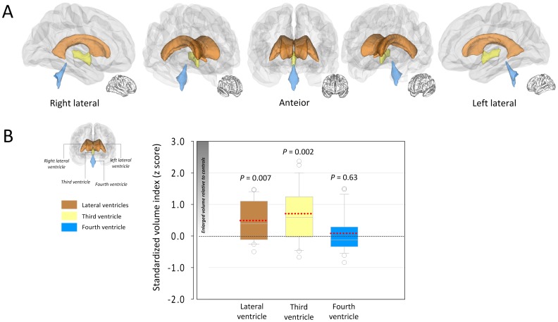
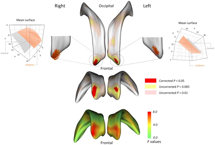
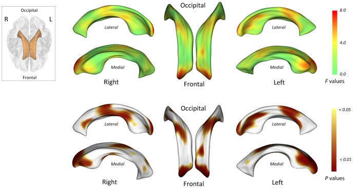
It is becoming increasingly evident that type 2 diabetes mellitus can have effects on global and regional brain morphology. Ventricular enlargement reflecting cerebral atrophy has been reported particularly in elderly type 2 diabetes patients. However, little is known about its timing through the disease course and morphological variability. Using the combined volumetric and advanced three-dimensional morphological approach, we identified differences in size and shape of the lateral ventricles between recent-onset type 2 diabetes patients and healthy individuals. High-resolution T1-weighted images were obtained from 23 type 2 diabetes patients whose illness duration was less than 1 year and 23 carefully matched healthy individuals. By volume measurement, we found enlarged lateral and third ventricles in type 2 diabetes patients, relative to healthy individuals (F(1,41 )= 7.96, P = 0.007; F(1,41) = 11.16, P = 0.002, respectively). Morphological analysis revealed that the expansion of lateral ventricles in the diabetic brain was prominent in the bilateral frontal horns. The current findings suggest that atrophic changes particularly of the anterior frontal lobe can occur as early as the first year after the clinical diagnosis of type 2 diabetes mellitus.
越来越明显的是,2 型糖尿病可能对全球和区域脑形态有影响。脑室扩大反映脑萎缩在老年 2 型糖尿病患者中尤为常见。然而,关于其在疾病过程中的时间和形态变化知之甚少。我们使用组合容积和先进的三维形态方法,在近期发病的 2 型糖尿病患者和健康个体之间发现了侧脑室大小和形状的差异。从病程不足 1 年的 23 名 2 型糖尿病患者和 23 名精心匹配的健康个体中获得了高分辨率 T1 加权图像。通过体积测量,我们发现与健康个体相比,2 型糖尿病患者的侧脑室和第三脑室扩大(F(1,41)=7.96,P=0.007;F(1,41)=11.16,P=0.002)。形态分析显示,糖尿病大脑中侧脑室的扩张在前额角两侧最为明显。目前的研究结果表明,在临床诊断 2 型糖尿病后的第一年,就可能出现前额叶的萎缩性变化。