Laboratory for Living Systems Engineering, Department of Biomedical Engineering, USC Viterbi School of Engineering, University of Southern California, Los Angeles, CA, 90089, USA.
Oxford Instruments Asylum Research, Santa Barbara, CA, 93117, USA.
Sci Rep. 2016 Jun 28;6:28855. doi: 10.1038/srep28855.
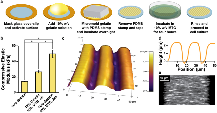
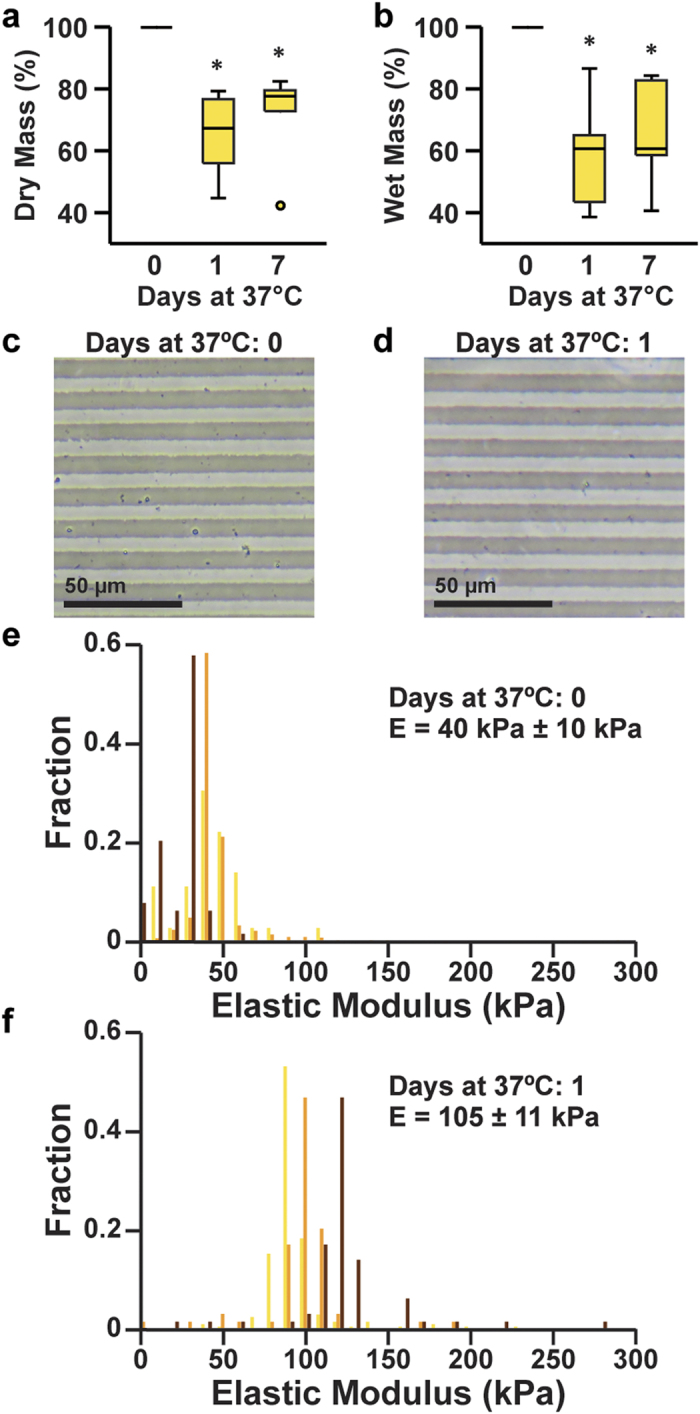
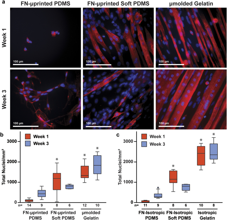
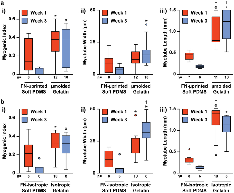
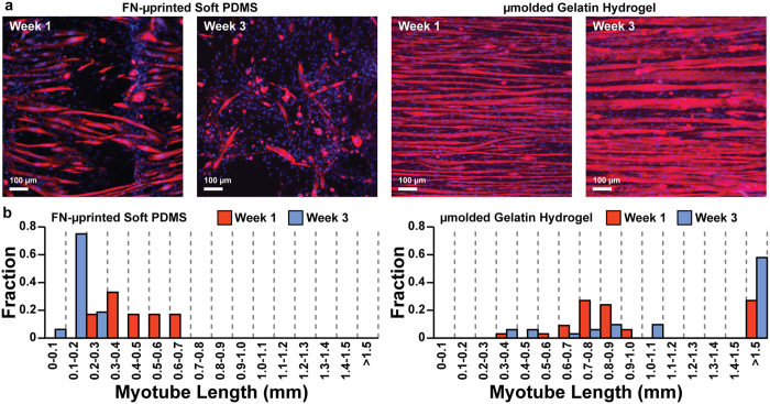
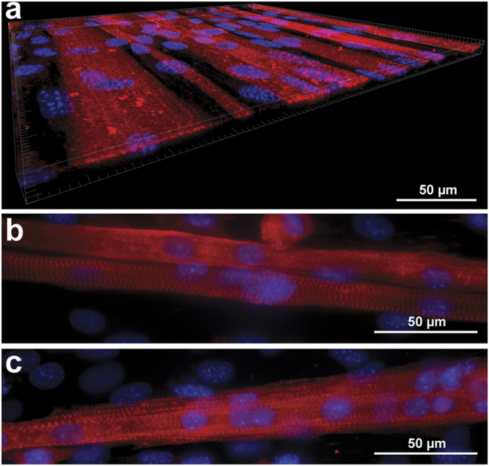
In vitro models of skeletal muscle are critically needed to elucidate disease mechanisms, identify therapeutic targets, and test drugs pre-clinically. However, culturing skeletal muscle has been challenging due to myotube delamination from synthetic culture substrates approximately one week after initiating differentiation from myoblasts. In this study, we successfully maintained aligned skeletal myotubes differentiated from C2C12 mouse skeletal myoblasts for three weeks by utilizing micromolded (μmolded) gelatin hydrogels as culture substrates, which we thoroughly characterized using atomic force microscopy (AFM). Compared to polydimethylsiloxane (PDMS) microcontact printed (μprinted) with fibronectin (FN), cell adhesion on gelatin hydrogel constructs was significantly higher one week and three weeks after initiating differentiation. Delamination from FN-μprinted PDMS precluded robust detection of myotubes. Compared to a softer blend of PDMS μprinted with FN, myogenic index, myotube width, and myotube length on μmolded gelatin hydrogels was similar one week after initiating differentiation. However, three weeks after initiating differentiation, these parameters were significantly higher on μmolded gelatin hydrogels compared to FN-μprinted soft PDMS constructs. Similar results were observed on isotropic versions of each substrate, suggesting that these findings are independent of substrate patterning. Our platform enables novel studies into skeletal muscle development and disease and chronic drug testing in vitro.
体外骨骼肌模型对于阐明疾病机制、确定治疗靶点以及在临床前测试药物至关重要。然而,由于骨骼肌从成肌细胞分化后大约一周就会从合成培养基质上分层,因此培养骨骼肌一直具有挑战性。在这项研究中,我们通过使用微成型(μmolded)明胶水凝胶作为培养基质,成功地将从 C2C12 小鼠骨骼肌成肌细胞分化而来的排列整齐的骨骼肌肌管维持了 3 周,我们使用原子力显微镜(AFM)对其进行了全面表征。与用纤维连接蛋白(FN)微接触印刷(μprinted)的聚二甲基硅氧烷(PDMS)相比,在开始分化一周和三周后,细胞在明胶水凝胶构建体上的黏附显著更高。FN-μprinted PDMS 的分层使肌管的稳健检测变得困难。与 FN-μprinted 较软的 PDMS 混合物相比,在开始分化一周后,μmolded 明胶水凝胶上的成肌指数、肌管宽度和肌管长度相似。然而,在开始分化三周后,这些参数在 μmolded 明胶水凝胶上明显高于 FN-μprinted 软 PDMS 构建体。在每个基质的各向同性版本上都观察到了类似的结果,这表明这些发现与基质图案化无关。我们的平台使人们能够对骨骼肌发育和疾病进行新的研究,并在体外进行慢性药物测试。