• 文献检索
  • 文档翻译
  • 深度研究
  • 学术资讯
  • Suppr Zotero 插件Zotero 插件
  • 邀请有礼
  • 套餐&价格
  • 历史记录
应用&插件
Suppr Zotero 插件Zotero 插件浏览器插件Mac 客户端Windows 客户端微信小程序
定价
高级版会员购买积分包购买API积分包
服务
文献检索文档翻译深度研究API 文档MCP 服务
关于我们
关于 Suppr公司介绍联系我们用户协议隐私条款
关注我们

Suppr 超能文献

核心技术专利:CN118964589B侵权必究
粤ICP备2023148730 号-1Suppr @ 2026

文献检索

告别复杂PubMed语法,用中文像聊天一样搜索,搜遍4000万医学文献。AI智能推荐,让科研检索更轻松。

立即免费搜索

文件翻译

保留排版,准确专业,支持PDF/Word/PPT等文件格式,支持 12+语言互译。

免费翻译文档

深度研究

AI帮你快速写综述,25分钟生成高质量综述,智能提取关键信息,辅助科研写作。

立即免费体验

在单细胞尺度上构建人类骨骼肌的 3D 体外模型。

Engineering a 3D in vitro model of human skeletal muscle at the single fiber scale.

机构信息

Industrial Engineering Department, University of Padova, Padova, Italy.

Venetian Institute of Molecular Medicine, Padova, Italy.

出版信息

PLoS One. 2020 May 6;15(5):e0232081. doi: 10.1371/journal.pone.0232081. eCollection 2020.

DOI:10.1371/journal.pone.0232081
PMID:32374763
原文链接:https://pmc.ncbi.nlm.nih.gov/articles/PMC7202609/
Abstract

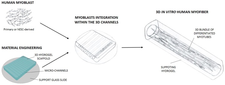
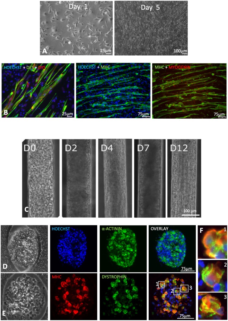
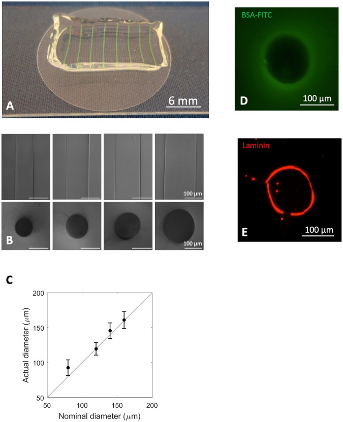
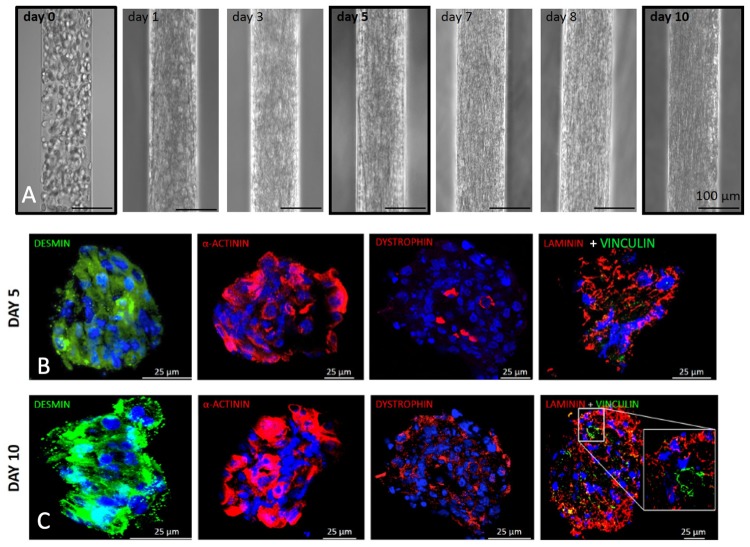
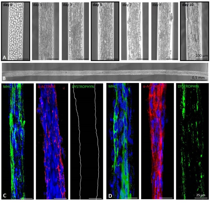
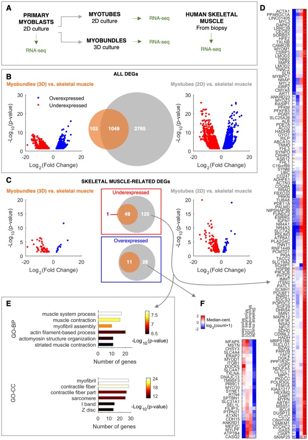
The reproduction of reliable in vitro models of human skeletal muscle is made harder by the intrinsic 3D structural complexity of this tissue. Here we coupled engineered hydrogel with 3D structural cues and specific mechanical properties to derive human 3D muscle constructs ("myobundles") at the scale of single fibers, by using primary myoblasts or myoblasts derived from embryonic stem cells. To this aim, cell culture was performed in confined, laminin-coated micrometric channels obtained inside a 3D hydrogel characterized by the optimal stiffness for skeletal muscle myogenesis. Primary myoblasts cultured in our 3D culture system were able to undergo myotube differentiation and maturation, as demonstrated by the proper expression and localization of key components of the sarcomere and sarcolemma. Such approach allowed the generation of human myobundles of ~10 mm in length and ~120 μm in diameter, showing spontaneous contraction 7 days after cell seeding. Transcriptome analyses showed higher similarity between 3D myobundles and skeletal signature, compared to that found between 2D myotubes and skeletal muscle, mainly resulting from expression in 3D myobundles of categories of genes involved in skeletal muscle maturation, including extracellular matrix organization. Moreover, imaging analyses confirmed that structured 3D culture system was conducive to differentiation/maturation also when using myoblasts derived from embryonic stem cells. In conclusion, our structured 3D model is a promising tool for modelling human skeletal muscle in healthy and diseases conditions.

摘要

人体骨骼肌的可靠体外模型的再现由于该组织固有的 3D 结构复杂性而变得更加困难。在这里,我们通过使用原代成肌细胞或胚胎干细胞衍生的成肌细胞,将工程水凝胶与 3D 结构线索和特定的机械性能相结合,在单个纤维的尺度上获得了人类 3D 肌肉构建体(“肌束”)。为此,细胞培养是在受限的、层粘连蛋白包被的微尺度通道内进行的,这些通道是在具有骨骼肌成肌作用最佳刚度的 3D 水凝胶内获得的。在我们的 3D 培养系统中培养的原代成肌细胞能够进行肌管分化和成熟,这可以通过肌节和肌膜关键成分的正确表达和定位来证明。这种方法允许生成约 10mm 长和 120μm 直径的人类肌束,在细胞接种后 7 天显示自发收缩。转录组分析表明,与 2D 肌管和骨骼肌相比,3D 肌束与骨骼肌的特征更为相似,这主要是由于 3D 肌束中表达了与骨骼肌成熟相关的基因类别,包括细胞外基质组织。此外,成像分析证实,结构 3D 培养系统有利于分化/成熟,即使使用源自胚胎干细胞的成肌细胞也是如此。总之,我们的结构化 3D 模型是在健康和疾病条件下模拟人类骨骼肌的有前途的工具。

https://cdn.ncbi.nlm.nih.gov/pmc/blobs/a1bb/7202609/a7526eac217a/pone.0232081.g006.jpg
https://cdn.ncbi.nlm.nih.gov/pmc/blobs/a1bb/7202609/2f7298d13fa6/pone.0232081.g001.jpg
https://cdn.ncbi.nlm.nih.gov/pmc/blobs/a1bb/7202609/c4fae57f046b/pone.0232081.g002.jpg
https://cdn.ncbi.nlm.nih.gov/pmc/blobs/a1bb/7202609/6f7afbfe2e8d/pone.0232081.g003.jpg
https://cdn.ncbi.nlm.nih.gov/pmc/blobs/a1bb/7202609/95600c091ba4/pone.0232081.g004.jpg
https://cdn.ncbi.nlm.nih.gov/pmc/blobs/a1bb/7202609/e96f43159058/pone.0232081.g005.jpg
https://cdn.ncbi.nlm.nih.gov/pmc/blobs/a1bb/7202609/a7526eac217a/pone.0232081.g006.jpg

相似文献

1
Engineering a 3D in vitro model of human skeletal muscle at the single fiber scale.在单细胞尺度上构建人类骨骼肌的 3D 体外模型。
PLoS One. 2020 May 6;15(5):e0232081. doi: 10.1371/journal.pone.0232081. eCollection 2020.
2
Electrical stimulation increases hypertrophy and metabolic flux in tissue-engineered human skeletal muscle.电刺激可增加组织工程化人骨骼肌的肥大和代谢通量。
Biomaterials. 2019 Apr;198:259-269. doi: 10.1016/j.biomaterials.2018.08.058. Epub 2018 Aug 31.
3
Prolonged Culture of Aligned Skeletal Myotubes on Micromolded Gelatin Hydrogels.在微成型明胶水凝胶上培养对齐的骨骼肌肌管延长时间。
Sci Rep. 2016 Jun 28;6:28855. doi: 10.1038/srep28855.
4
Cell Density and Joint microRNA-133a and microRNA-696 Inhibition Enhance Differentiation and Contractile Function of Engineered Human Skeletal Muscle Tissues.细胞密度以及关节微小RNA - 133a和微小RNA - 696的抑制可增强工程化人骨骼肌组织的分化和收缩功能。
Tissue Eng Part A. 2016 Apr;22(7-8):573-83. doi: 10.1089/ten.TEA.2015.0359.
5
Microfluidic-enhanced 3D bioprinting of aligned myoblast-laden hydrogels leads to functionally organized myofibers in vitro and in vivo.微流控增强的 3D 生物打印技术可将负载成肌细胞的水凝胶排列整齐,从而在体外和体内构建具有功能组织结构的肌纤维。
Biomaterials. 2017 Jul;131:98-110. doi: 10.1016/j.biomaterials.2017.03.026. Epub 2017 Mar 23.
6
Mesenchymal stem cells and myoblast differentiation under HGF and IGF-1 stimulation for 3D skeletal muscle tissue engineering.用于3D骨骼肌组织工程的间充质干细胞和成肌细胞在HGF和IGF-1刺激下的分化
BMC Cell Biol. 2017 Feb 28;18(1):15. doi: 10.1186/s12860-017-0131-2.
7
Myoblast maturity on aligned microfiber bundles at the onset of strain application impacts myogenic outcomes.在开始施加应变时,位于取向微纤维束上的成肌细胞成熟程度会影响成肌效果。
Acta Biomater. 2019 Aug;94:232-242. doi: 10.1016/j.actbio.2019.06.024. Epub 2019 Jun 15.
8
Fabrication of contractile skeletal muscle tissues using directly converted myoblasts from human fibroblasts.利用源自人成纤维细胞的直接转化肌母细胞来构建收缩性骨骼肌组织。
J Biosci Bioeng. 2020 May;129(5):632-637. doi: 10.1016/j.jbiosc.2019.11.013. Epub 2019 Dec 16.
9
Satellite cells delivered in their niche efficiently generate functional myotubes in three-dimensional cell culture.在三维细胞培养中,位于其龛位中的卫星细胞能够有效地生成具有功能的肌管。
PLoS One. 2018 Sep 17;13(9):e0202574. doi: 10.1371/journal.pone.0202574. eCollection 2018.
10
Three-dimensional co-culture of C2C12/PC12 cells improves skeletal muscle tissue formation and function.C2C12/PC12 细胞的三维共培养可改善骨骼肌组织的形成和功能。
J Tissue Eng Regen Med. 2017 Feb;11(2):582-595. doi: 10.1002/term.1956. Epub 2014 Nov 13.

引用本文的文献

1
Fundamental study on structural formation, amino acids and nucleotide-related compounds of cultivated meat from 3D-cultured pig muscle stem cells.基于3D培养猪肌肉干细胞的培养肉的结构形成、氨基酸及核苷酸相关化合物的基础研究
Food Sci Biotechnol. 2024 Dec 17;34(2):457-469. doi: 10.1007/s10068-024-01793-9. eCollection 2025 Jan.
2
Multitasking muscle: engineering iPSC-derived myogenic progenitors to do more.多功能肌肉:工程化诱导多能干细胞衍生的成肌祖细胞以发挥更多功能。
Front Cell Dev Biol. 2025 Jan 22;12:1526635. doi: 10.3389/fcell.2024.1526635. eCollection 2024.
3
Near Infrared-Mediated Intracellular NADH Delivery Strengthens Mitochondrial Function and Stability in Muscle Dysfunction Model.

本文引用的文献

1
3D Bioprinting in Skeletal Muscle Tissue Engineering.三维生物打印在骨骼肌组织工程中的应用。
Small. 2019 Jun;15(24):e1805530. doi: 10.1002/smll.201805530. Epub 2019 Apr 23.
2
Engineering an Environment for the Study of Fibrosis: A 3D Human Muscle Model with Endothelium Specificity and Endomysium.工程化纤维化研究环境:具有血管内皮特异性和肌内膜的 3D 人类肌肉模型。
Cell Rep. 2018 Dec 26;25(13):3858-3868.e4. doi: 10.1016/j.celrep.2018.11.092.
3
Electrical stimulation increases hypertrophy and metabolic flux in tissue-engineered human skeletal muscle.
近红外介导的细胞内烟酰胺腺嘌呤二核苷酸递送增强肌肉功能障碍模型中的线粒体功能和稳定性
Adv Sci (Weinh). 2025 Mar;12(12):e2415303. doi: 10.1002/advs.202415303. Epub 2025 Jan 31.
4
Endothelial-mesenchymal transition in skeletal muscle: Opportunities and challenges from 3D microphysiological systems.骨骼肌中的内皮-间充质转化:来自3D微生理系统的机遇与挑战
Bioeng Transl Med. 2024 Jan 29;9(5):e10644. doi: 10.1002/btm2.10644. eCollection 2024 Sep.
5
Differential Fatty Acid Response of Resident Macrophages in Human Skeletal Muscle Fiber and Intermuscular Adipose Tissue.人类骨骼肌纤维和肌间脂肪组织中驻留巨噬细胞的脂肪酸反应差异。
Int J Mol Sci. 2024 Oct 5;25(19):10722. doi: 10.3390/ijms251910722.
6
Advancing insights into microgravity induced muscle changes using Caenorhabditis elegans as a model organism.以秀丽隐杆线虫作为模式生物,深入了解微重力引起的肌肉变化。
NPJ Microgravity. 2024 Jul 26;10(1):79. doi: 10.1038/s41526-024-00418-z.
7
Nano-biomaterials and advanced fabrication techniques for engineering skeletal muscle tissue constructs in regenerative medicine.用于再生医学中工程化骨骼肌组织构建体的纳米生物材料和先进制造技术。
Nano Converg. 2023 Oct 21;10(1):48. doi: 10.1186/s40580-023-00398-y.
8
Enhanced Maturation of 3D Bioprinted Skeletal Muscle Tissue Constructs Encapsulating Soluble Factor-Releasing Microparticles.增强型三维生物打印骨骼肌组织构建体的成熟,构建体中封装了可溶性因子释放微球。
Macromol Biosci. 2023 Dec;23(12):e2300276. doi: 10.1002/mabi.202300276. Epub 2023 Aug 17.
9
Advancements in the Use of Hydrogels for Regenerative Medicine: Properties and Biomedical Applications.水凝胶在再生医学中的应用进展:性质与生物医学应用
Int J Biomater. 2022 Nov 7;2022:3606765. doi: 10.1155/2022/3606765. eCollection 2022.
10
Biomaterials for Regenerative Medicine in Italy: Brief State of the Art of the Principal Research Centers.意大利再生医学用生物材料:主要研究中心的简要现状概述。
Int J Mol Sci. 2022 Jul 26;23(15):8245. doi: 10.3390/ijms23158245.
电刺激可增加组织工程化人骨骼肌的肥大和代谢通量。
Biomaterials. 2019 Apr;198:259-269. doi: 10.1016/j.biomaterials.2018.08.058. Epub 2018 Aug 31.
4
3D Bioprinted Human Skeletal Muscle Constructs for Muscle Function Restoration.3D 生物打印的人类骨骼肌构建体用于肌肉功能恢复。
Sci Rep. 2018 Aug 17;8(1):12307. doi: 10.1038/s41598-018-29968-5.
5
Decellularized Tissue for Muscle Regeneration.脱细胞组织用于肌肉再生。
Int J Mol Sci. 2018 Aug 14;19(8):2392. doi: 10.3390/ijms19082392.
6
Decellularised skeletal muscles allow functional muscle regeneration by promoting host cell migration.去细胞化的骨骼肌通过促进宿主细胞迁移来实现功能性肌肉再生。
Sci Rep. 2018 May 30;8(1):8398. doi: 10.1038/s41598-018-26371-y.
7
Current Progress and Challenges for Skeletal Muscle Differentiation from Human Pluripotent Stem Cells Using Transgene-Free Approaches.使用无转基因方法从人多能干细胞分化骨骼肌的当前进展与挑战
Stem Cells Int. 2018 Apr 11;2018:6241681. doi: 10.1155/2018/6241681. eCollection 2018.
8
In Vitro Tissue-Engineered Skeletal Muscle Models for Studying Muscle Physiology and Disease.用于研究肌肉生理学和疾病的体外组织工程化骨骼肌模型。
Adv Healthc Mater. 2018 Aug;7(15):e1701498. doi: 10.1002/adhm.201701498. Epub 2018 Apr 25.
9
Three-Dimensional Human iPSC-Derived Artificial Skeletal Muscles Model Muscular Dystrophies and Enable Multilineage Tissue Engineering.三维人诱导多能干细胞衍生的人工骨骼肌模型模拟肌肉疾病并实现多谱系组织工程。
Cell Rep. 2018 Apr 17;23(3):899-908. doi: 10.1016/j.celrep.2018.03.091.
10
Hydrogel biomaterials and their therapeutic potential for muscle injuries and muscular dystrophies.水凝胶生物材料及其在肌肉损伤和肌肉疾病治疗中的潜力。
J R Soc Interface. 2018 Jan;15(138). doi: 10.1098/rsif.2017.0380.