Zhao Yuguang, Ren Jingshan, Harlos Karl, Jones Daniel M, Zeltina Antra, Bowden Thomas A, Padilla-Parra Sergi, Fry Elizabeth E, Stuart David I
Division of Structural Biology, University of Oxford, The Henry Wellcome Building for Genomic Medicine, Headington, Oxford, OX3 7BN, UK.
Cellular Imaging Core, Wellcome Trust Centre for Human Genetics, University of Oxford, Oxford, UK.
Nature. 2016 Jul 7;535(7610):169-172. doi: 10.1038/nature18615. Epub 2016 Jun 29.
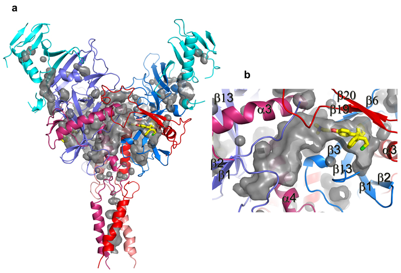
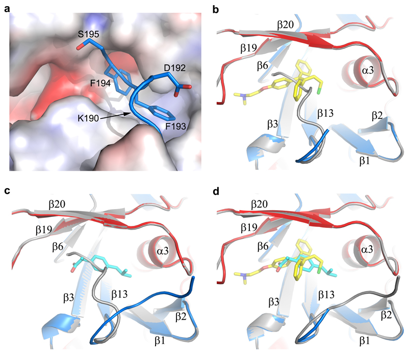
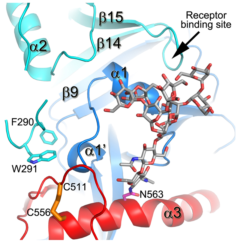
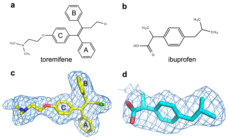
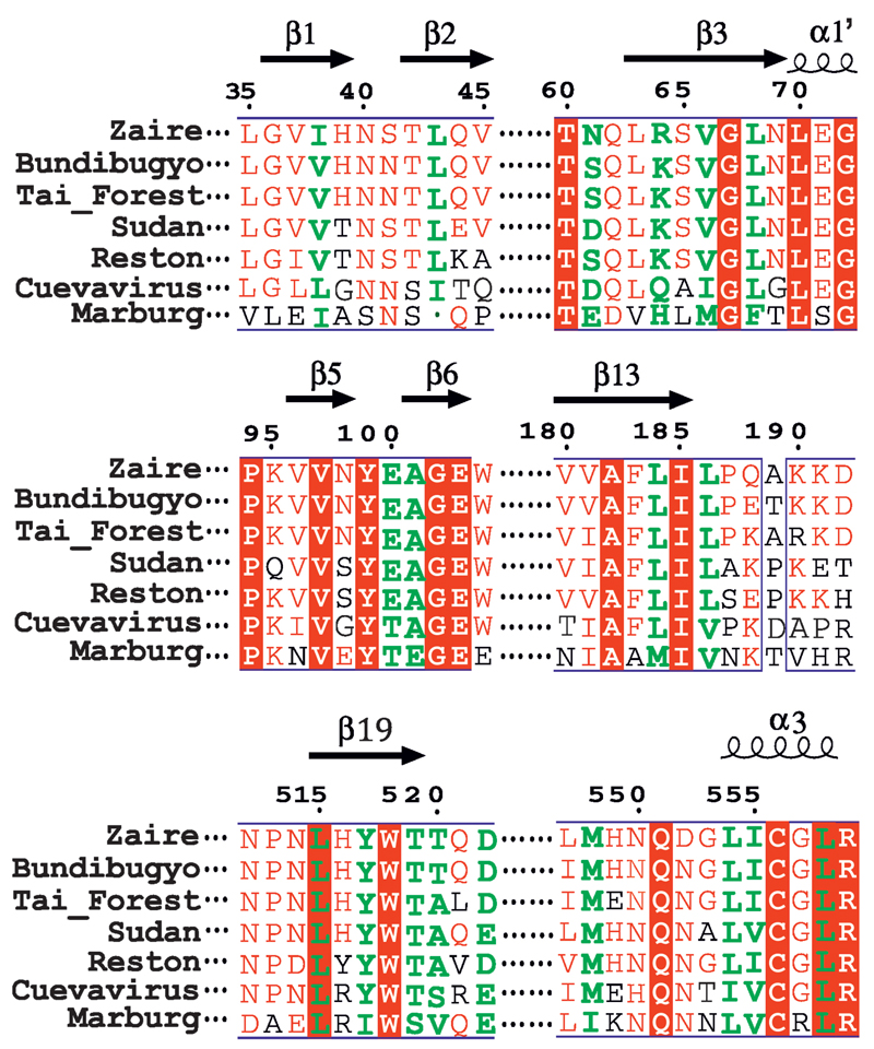
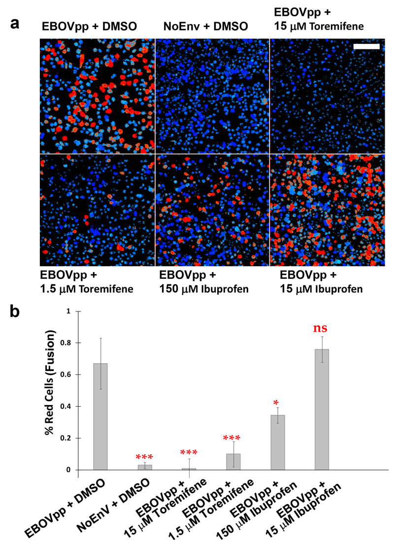
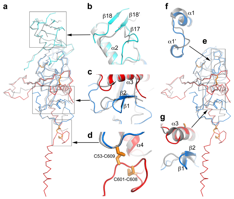
Ebola viruses (EBOVs) are responsible for repeated outbreaks of fatal infections, including the recent deadly epidemic in West Africa. There are currently no approved therapeutic drugs or vaccines for the disease. EBOV has a membrane envelope decorated by trimers of a glycoprotein (GP, cleaved by furin to form GP1 and GP2 subunits), which is solely responsible for host cell attachment, endosomal entry and membrane fusion. GP is thus a primary target for the development of antiviral drugs. Here we report the first, to our knowledge, unliganded structure of EBOV GP, and high-resolution complexes of GP with the anticancer drug toremifene and the painkiller ibuprofen. The high-resolution apo structure gives a more complete and accurate picture of the molecule, and allows conformational changes introduced by antibody and receptor binding to be deciphered. Unexpectedly, both toremifene and ibuprofen bind in a cavity between the attachment (GP1) and fusion (GP2) subunits at the entrance to a large tunnel that links with equivalent tunnels from the other monomers of the trimer at the three-fold axis. Protein–drug interactions with both GP1 and GP2 are predominately hydrophobic. Residues lining the binding site are highly conserved among filoviruses except Marburg virus (MARV), suggesting that MARV may not bind these drugs. Thermal shift assays show up to a 14 °C decrease in the protein melting temperature after toremifene binding, while ibuprofen has only a marginal effect and is a less potent inhibitor. These results suggest that inhibitor binding destabilizes GP and triggers premature release of GP2, thereby preventing fusion between the viral and endosome membranes. Thus, these complex structures reveal the mechanism of inhibition and may guide the development of more powerful anti-EBOV drugs.
埃博拉病毒(EBOV)导致了多次致命感染的爆发,包括近期在西非发生的致命疫情。目前该疾病尚无获批的治疗药物或疫苗。EBOV具有一层包膜,包膜上装饰有糖蛋白(GP,被弗林蛋白酶切割形成GP1和GP2亚基)三聚体,该糖蛋白单独负责宿主细胞附着、内体进入和膜融合。因此,GP是抗病毒药物开发的主要靶点。在此,据我们所知,我们报道了EBOV GP的首个无配体结构,以及GP与抗癌药物托瑞米芬和止痛药布洛芬的高分辨率复合物结构。高分辨率的无配体结构给出了该分子更完整、准确的图像,并能解读由抗体和受体结合引入的构象变化。出乎意料的是,托瑞米芬和布洛芬都结合在附着亚基(GP1)和融合亚基(GP2)之间的一个腔中,该腔位于一条大通道的入口处,该通道与三聚体其他单体在三重轴处的等效通道相连。与GP1和GP2的蛋白质 - 药物相互作用主要是疏水作用。除马尔堡病毒(MARV)外,在丝状病毒中,结合位点周围的残基高度保守,这表明MARV可能不结合这些药物。热迁移分析表明,托瑞米芬结合后蛋白质解链温度降低高达14°C,而布洛芬只有轻微影响,是一种效力较弱的抑制剂。这些结果表明抑制剂结合使GP不稳定并触发GP2的过早释放,从而阻止病毒膜与内体膜之间的融合。因此,这些复杂结构揭示了抑制机制,并可能指导更有效的抗EBOV药物的开发。