Payen Thomas, Palermo Carmine F, Sastra Stephen A, Chen Hong, Han Yang, Olive Kenneth P, Konofagou Elisa E
Biomedical Engineering, Columbia University, USA.
Phys Med Biol. 2016 Aug 7;61(15):5741-54. doi: 10.1088/0031-9155/61/15/5741. Epub 2016 Jul 12.
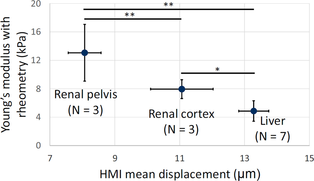
Recently, ultrasonic imaging of soft tissue mechanics has been increasingly studied to image otherwise undetectable pathologies. However, many underlying mechanisms of tissue stiffening remain unknown, requiring small animal studies and adapted elasticity mapping techniques. Harmonic motion imaging (HMI) assesses tissue viscoelasticity by inducing localized oscillation from a periodic acoustic radiation force. The objective of this study was to evaluate the feasibility of HMI for in vivo elasticity mapping of abdominal organs in small animals. Pathological cases, i.e. chronic pancreatitis and pancreatic cancer, were also studied in vivo to assess the capability of HMI for detection of the change in mechanical properties. A 4.5 MHz focused ultrasound transducer (FUS) generated an amplitude-modulated beam resulting in 50 Hz harmonic tissue oscillations at its focus. Axial tissue displacement was estimated using 1D-cross-correlation of RF signals acquired with a 7.8 MHz diagnostic transducer confocally aligned with the FUS. In vitro results in canine liver and kidney showed the correlation between HMI displacement and Young's moduli measured by rheometry compression testing. HMI was capable of providing reproducible elasticity maps of the mouse abdominal region in vivo allowing the identification of, from stiffest to softest, the murine kidney, pancreas, liver, and spleen. Finally, pancreata affected by pancreatitis and pancreatic cancer showed HMI displacements 1.7 and 2.2 times lower than in the control case, respectively, indicating higher stiffness. The HMI displacement amplitude was correlated with the extent of fibrosis as well as detecting the very onset of stiffening even before fibrosis could be detected on H&E. This work shows that HMI can produce reliable elasticity maps of mouse abdominal region in vivo, thus providing a potentially critical tool to assess pathologies affecting organ elasticity.
最近,软组织力学的超声成像技术越来越多地被用于对原本无法检测到的病变进行成像。然而,组织硬化的许多潜在机制仍然未知,这需要进行小动物研究并采用适应性弹性映射技术。谐波运动成像(HMI)通过周期性声辐射力诱导局部振荡来评估组织的粘弹性。本研究的目的是评估HMI在小动物体内对腹部器官进行弹性映射的可行性。还对病理病例,即慢性胰腺炎和胰腺癌进行了体内研究,以评估HMI检测机械性能变化的能力。一个4.5MHz的聚焦超声换能器(FUS)产生一个调幅波束,在其焦点处产生50Hz的谐波组织振荡。使用与FUS共焦对准的7.8MHz诊断换能器采集的RF信号的一维互相关来估计轴向组织位移。犬肝脏和肾脏的体外结果显示了HMI位移与通过流变学压缩测试测量的杨氏模量之间的相关性。HMI能够在体内提供小鼠腹部区域的可重复弹性图,从而能够从最硬到最软识别出小鼠的肾脏、胰腺、肝脏和脾脏。最后,受胰腺炎和胰腺癌影响的胰腺显示HMI位移分别比对照病例低1.7倍和2.2倍,表明硬度更高。HMI位移幅度与纤维化程度相关,甚至在苏木精-伊红染色(H&E)检测到纤维化之前就能检测到硬化的最初阶段。这项工作表明,HMI能够在体内生成小鼠腹部区域可靠的弹性图,从而为评估影响器官弹性的病变提供了一个潜在的关键工具。