Wanek Justin, Blair Norman P, Chau Felix Y, Lim Jennifer I, Leiderman Yannek I, Shahidi Mahnaz
Invest Ophthalmol Vis Sci. 2016 Jul 1;57(9):OCT341-7. doi: 10.1167/iovs.15-18715.
This article reports a method for en face optical coherence tomography (OCT) imaging and quantitative assessment of alterations in both thickness and reflectance of individual retinal layers at different stages of diabetic retinopathy (DR).
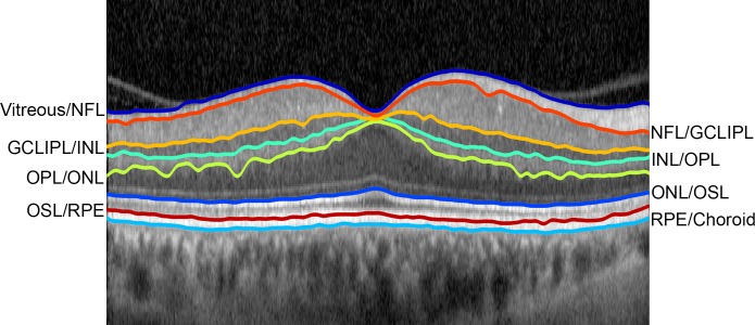
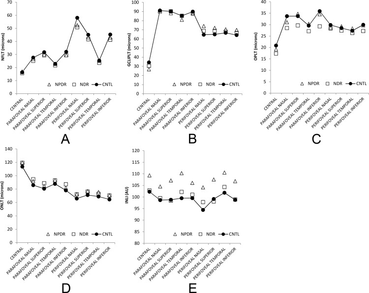
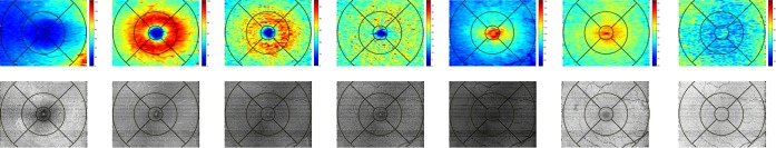
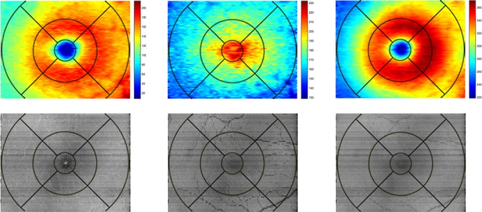
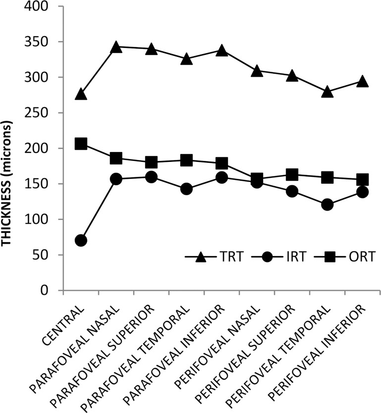
High-density OCT raster volume scans were acquired in 29 diabetic subjects divided into no DR (NDR) or non-proliferative DR (NPDR) groups and 22 control subjects (CNTL). A customized image segmentation method identified eight retinal layer interfaces and generated en face thickness maps and reflectance images for nerve fiber layer (NFL), ganglion cell and inner plexiform layers (GCLIPL), inner nuclear layer (INL), outer plexiform layer (OPL), outer nuclear layer (ONL), photoreceptor outer segment layer (OSL), and retinal pigment epithelium (RPE). Mean thickness and intensity values were calculated in nine macular subfields for each retinal layer.
En face thickness maps and reflectance images of retinal layers in CNTL subjects corresponded to normal retinal anatomy. Total retinal thickness correlated negatively with age in nasal subfields (R ≤-0.31; P ≤ 0.03, N = 51). In NDR subjects, NFL and OPL thickness were decreased (P = 0.05), and ONL thickness was increased (P = 0.04) compared to CNTL. In NPDR subjects, GCLIPL thickness was increased in perifoveal subfields (P< 0.05) and INL intensity was higher in all macular subfields (P = 0.04) compared to CNTL.
Depth and spatially resolved retinal thickness and reflectance measurements are potential biomarkers for assessment and monitoring of DR.
本文报告一种用于糖尿病性视网膜病变(DR)不同阶段个体视网膜各层厚度和反射率变化的表面光学相干断层扫描(OCT)成像及定量评估方法。
对29例糖尿病患者(分为无糖尿病性视网膜病变(NDR)组或非增殖性糖尿病性视网膜病变(NPDR)组)和22例对照受试者(CNTL)进行高密度OCT光栅容积扫描。一种定制的图像分割方法识别出八个视网膜层界面,并生成神经纤维层(NFL)、神经节细胞和内丛状层(GCLIPL)、内核层(INL)、外丛状层(OPL)、外核层(ONL)、光感受器外段层(OSL)和视网膜色素上皮(RPE)的表面厚度图和反射率图像。计算每个视网膜层在九个黄斑子区域的平均厚度和强度值。
CNTL受试者视网膜层的表面厚度图和反射率图像与正常视网膜解剖结构相符。鼻侧子区域的总视网膜厚度与年龄呈负相关(R≤-0.31;P≤0.03,N = 51)。与CNTL相比,NDR受试者的NFL和OPL厚度降低(P = 0.05),ONL厚度增加(P = 0.04)。与CNTL相比,NPDR受试者的中央凹周围子区域GCLIPL厚度增加(P<0.05),所有黄斑子区域的INL强度更高(P = 0.04)。
深度和空间分辨的视网膜厚度及反射率测量是评估和监测糖尿病性视网膜病变的潜在生物标志物。