• 文献检索
  • 文档翻译
  • 深度研究
  • 学术资讯
  • Suppr Zotero 插件Zotero 插件
  • 邀请有礼
  • 套餐&价格
  • 历史记录
应用&插件
Suppr Zotero 插件Zotero 插件浏览器插件Mac 客户端Windows 客户端微信小程序
定价
高级版会员购买积分包购买API积分包
服务
文献检索文档翻译深度研究API 文档MCP 服务
关于我们
关于 Suppr公司介绍联系我们用户协议隐私条款
关注我们

Suppr 超能文献

核心技术专利:CN118964589B侵权必究
粤ICP备2023148730 号-1Suppr @ 2026

文献检索

告别复杂PubMed语法,用中文像聊天一样搜索,搜遍4000万医学文献。AI智能推荐,让科研检索更轻松。

立即免费搜索

文件翻译

保留排版,准确专业,支持PDF/Word/PPT等文件格式,支持 12+语言互译。

免费翻译文档

深度研究

AI帮你快速写综述,25分钟生成高质量综述,智能提取关键信息,辅助科研写作。

立即免费体验

视神经病变中视觉注视偏心度和不稳定性模式及其与视网膜神经节细胞层厚度的空间关系。

The Pattern of Visual Fixation Eccentricity and Instability in Optic Neuropathy and Its Spatial Relationship to Retinal Ganglion Cell Layer Thickness.

作者信息

Mallery Robert M, Poolman Pieter, Thurtell Matthew J, Wang Jui-Kai, Garvin Mona K, Ledolter Johannes, Kardon Randy H

机构信息

Department of Neurology, Brigham and Women's Hospital, Boston, Massachusetts, United States 2Department of Ophthalmology, Massachusetts Eye and Ear Infirmary, Boston, Massachusetts, United States 3Department of Ophthalmology and Visual Sciences, Universit.

Iowa City VA Center for the Prevention and Treatment of Visual Loss, Iowa City, Iowa, United States.

出版信息

Invest Ophthalmol Vis Sci. 2016 Jul 1;57(9):OCT429-37. doi: 10.1167/iovs.15-18916.

DOI:10.1167/iovs.15-18916
PMID:27409502
原文链接:https://pmc.ncbi.nlm.nih.gov/articles/PMC4968926/
Abstract

PURPOSE

The purpose of this study was to assess whether clinically useful measures of fixation instability and eccentricity can be derived from retinal tracking data obtained during optical coherence tomography (OCT) in patients with optic neuropathy (ON) and to develop a method for relating fixation to the retinal ganglion cell complex (GCC) thickness.

METHODS

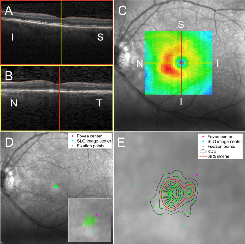
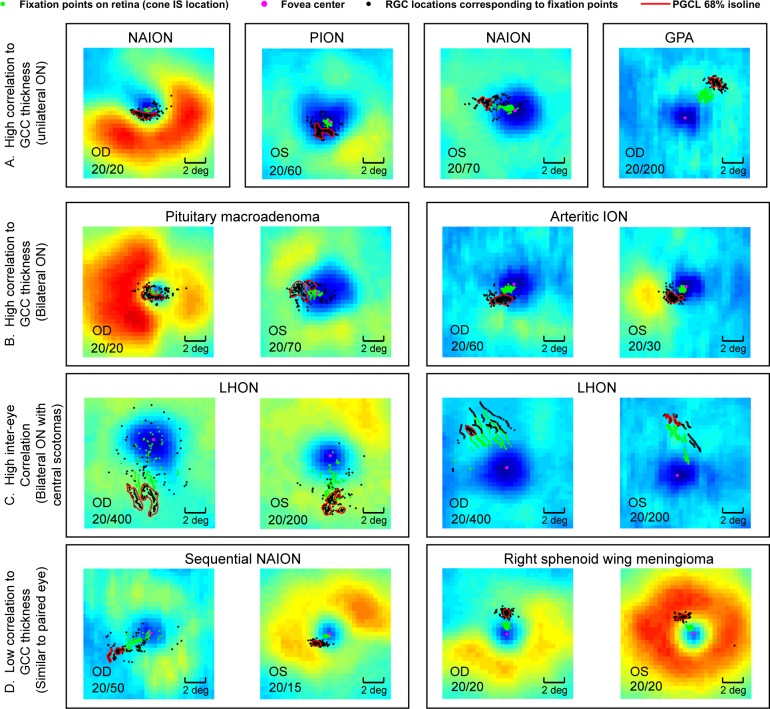
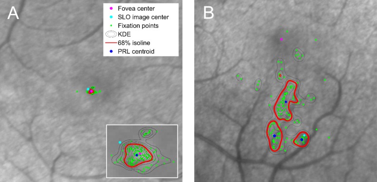
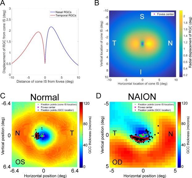
Twenty-nine patients with ON underwent macular volume OCT with 30 seconds of confocal scanning laser ophthalmoscope (cSLO)-based eye tracking during fixation. Kernel density estimation quantified fixation instability and fixation eccentricity from the distribution of fixation points on the retina. Preferred ganglion cell layer loci (PGCL) and their relationship to the GCC thickness map were derived, accounting for radial displacement of retinal ganglion cell soma from their corresponding cones.

RESULTS

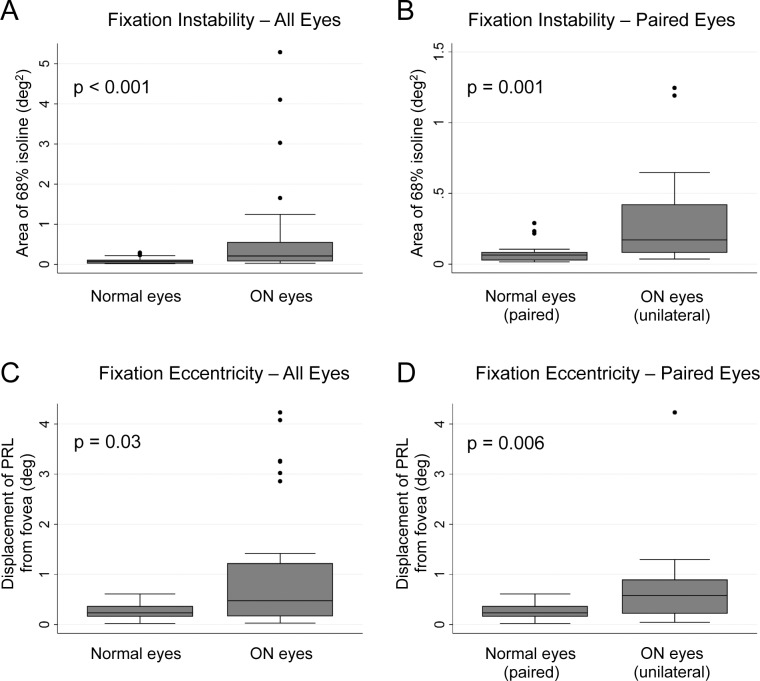
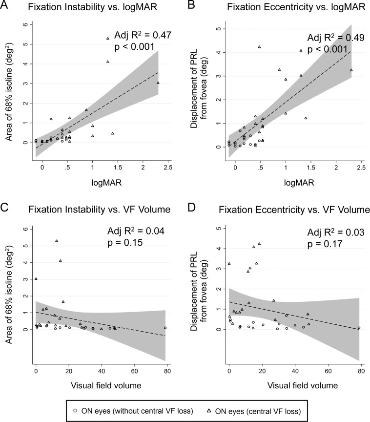
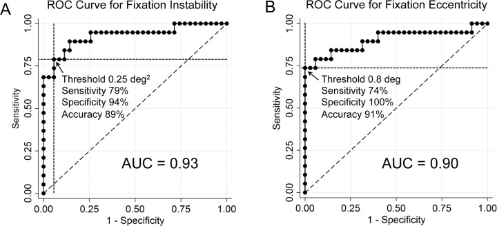
Fixation instability was increased in ON eyes (0.21 deg2) compared with normal eyes (0.06982 deg2; P < 0.001), and fixation eccentricity was increased in ON eyes (0.48°) compared with normal eyes (0.24°; P = 0.03). Fixation instability and eccentricity each correlated moderately with logMAR acuity and were highly predictive of central visual field loss. Twenty-six of 35 ON eyes had PGCL skewed toward local maxima of the GCC thickness map. Patients with bilateral dense central scotomas had PGCL in homonymous retinal locations with respect to the fovea.

CONCLUSIONS

Fixation instability and eccentricity measures obtained during cSLO-OCT assess the function of perifoveal retinal elements and predict central visual field loss in patients with ON. A model relating fixation to the GCC thickness map offers a method to assess the structure-function relationship between fixation and areas of preserved GCC in patients with ON.

摘要

目的

本研究旨在评估能否从视神经病变(ON)患者的光学相干断层扫描(OCT)过程中获得的视网膜追踪数据中得出临床上有用的注视不稳定和偏心度测量值,并开发一种将注视与视网膜神经节细胞复合体(GCC)厚度相关联的方法。

方法

29例ON患者在注视期间接受了黄斑容积OCT检查,并使用基于共焦扫描激光检眼镜(cSLO)的眼动追踪技术进行了30秒的扫描。核密度估计从视网膜上注视点的分布量化了注视不稳定和注视偏心度。得出了首选神经节细胞层位点(PGCL)及其与GCC厚度图的关系,同时考虑了视网膜神经节细胞胞体相对于其相应视锥细胞的径向位移。

结果

与正常眼(0.06982 deg2;P < 0.001)相比,ON患者患眼的注视不稳定增加(0.21 deg2),与正常眼(0.24°;P = 0.03)相比,ON患者患眼的注视偏心度增加(0.48°)。注视不稳定和偏心度均与logMAR视力呈中度相关,并能高度预测中心视野缺损。35只ON患眼中有26只的PGCL偏向GCC厚度图的局部最大值。双侧致密中心暗点的患者在相对于中央凹的同名视网膜位置有PGCL。

结论

在cSLO-OCT期间获得的注视不稳定和偏心度测量值可评估中心凹周围视网膜元件的功能,并预测ON患者的中心视野缺损。一种将注视与GCC厚度图相关联的模型提供了一种方法,用于评估ON患者注视与保留GCC区域之间的结构-功能关系。

https://cdn.ncbi.nlm.nih.gov/pmc/blobs/9172/4968926/3d1133a5df83/i1552-5783-57-9-OCT429-f07.jpg
https://cdn.ncbi.nlm.nih.gov/pmc/blobs/9172/4968926/e2ba1d1fdbaa/i1552-5783-57-9-OCT429-f01.jpg
https://cdn.ncbi.nlm.nih.gov/pmc/blobs/9172/4968926/6274f2c1f921/i1552-5783-57-9-OCT429-f02.jpg
https://cdn.ncbi.nlm.nih.gov/pmc/blobs/9172/4968926/3376f9e9fa79/i1552-5783-57-9-OCT429-f03.jpg
https://cdn.ncbi.nlm.nih.gov/pmc/blobs/9172/4968926/10dabeb02768/i1552-5783-57-9-OCT429-f04.jpg
https://cdn.ncbi.nlm.nih.gov/pmc/blobs/9172/4968926/685b76d6ab7e/i1552-5783-57-9-OCT429-f05.jpg
https://cdn.ncbi.nlm.nih.gov/pmc/blobs/9172/4968926/eb96bdc12d7e/i1552-5783-57-9-OCT429-f06.jpg
https://cdn.ncbi.nlm.nih.gov/pmc/blobs/9172/4968926/3d1133a5df83/i1552-5783-57-9-OCT429-f07.jpg

相似文献

1
The Pattern of Visual Fixation Eccentricity and Instability in Optic Neuropathy and Its Spatial Relationship to Retinal Ganglion Cell Layer Thickness.视神经病变中视觉注视偏心度和不稳定性模式及其与视网膜神经节细胞层厚度的空间关系。
Invest Ophthalmol Vis Sci. 2016 Jul 1;57(9):OCT429-37. doi: 10.1167/iovs.15-18916.
2
Comparison of retinal nerve fiber layer imaging by spectral domain optical coherence tomography and scanning laser ophthalmoscopy.频域光学相干断层扫描和扫描激光检眼镜对视神经纤维层成像的比较。
Ophthalmology. 2011 Nov;118(11):2196-202. doi: 10.1016/j.ophtha.2011.03.035. Epub 2011 Jul 16.
3
Difference in correspondence between visual field defect and inner macular layer thickness measured using three types of spectral-domain OCT instruments.使用三种类型的光谱域光学相干断层扫描(OCT)仪器测量时,视野缺损与黄斑内层厚度之间的对应差异。
Jpn J Ophthalmol. 2015 Jan;59(1):55-64. doi: 10.1007/s10384-014-0355-z. Epub 2014 Nov 7.
4
Retinal nerve fiber layer and ganglion cell complex thicknesses measured with spectral-domain optical coherence tomography in eyes with no light perception due to nonglaucomatous optic neuropathy.在因非青光眼性视神经病变而无光感的眼睛中,使用光谱域光学相干断层扫描测量视网膜神经纤维层和神经节细胞复合体厚度。
Jpn J Ophthalmol. 2015 Jul;59(4):230-5. doi: 10.1007/s10384-015-0386-0. Epub 2015 May 13.
5
Detection of macular ganglion cell loss in preperimetric glaucoma patients with localized retinal nerve fibre defects by spectral-domain optical coherence tomography.利用频域光学相干断层扫描检测局限性视网膜神经纤维层缺损的周边视野正常青光眼患者的黄斑神经节细胞丢失。
Clin Exp Ophthalmol. 2013 Dec;41(9):870-80. doi: 10.1111/ceo.12142. Epub 2013 Jul 5.
6
Alterations of the outer retina in non-arteritic anterior ischaemic optic neuropathy detected using spectral-domain optical coherence tomography.使用频域光学相干断层扫描检测非动脉炎性前部缺血性视神经病变中外层视网膜的改变。
Clin Exp Ophthalmol. 2017 Jul;45(5):496-508. doi: 10.1111/ceo.12914. Epub 2017 Mar 9.
7
Retinal nerve fiber layer imaging with spectral-domain optical coherence tomography a study on diagnostic agreement with Heidelberg Retinal Tomograph.频域光学相干断层扫描视网膜神经纤维层成像-海德堡视网膜断层扫描仪诊断符合度的研究。
Ophthalmology. 2010 Feb;117(2):267-74. doi: 10.1016/j.ophtha.2009.06.061. Epub 2009 Dec 6.
8
Comparison of retinal nerve fiber layer thickness values using Stratus Optical Coherence Tomography and Heidelberg Retina Tomograph-III.使用Stratus光学相干断层扫描和海德堡视网膜断层扫描仪III比较视网膜神经纤维层厚度值。
J Glaucoma. 2009 Sep;18(7):528-34. doi: 10.1097/IJG.0b013e318193c29f.
9
Comparison of confocal scanning laser ophthalmoscopy, scanning laser polarimetry and optical coherence tomography to discriminate ocular hypertension and glaucoma at an early stage.共焦扫描激光眼底镜、扫描激光偏振仪和光学相干断层扫描在早期鉴别高眼压症和青光眼的比较
Graefes Arch Clin Exp Ophthalmol. 2006 Jan;244(1):58-68. doi: 10.1007/s00417-005-0029-0. Epub 2005 Jul 26.
10
Correlation of localized glaucomatous visual field defects and spectral domain optical coherence tomography retinal nerve fiber layer thinning using a modified structure-function map for OCT.使用改良的光学相干断层扫描(OCT)结构-功能图对局限性青光眼视野缺损与光谱域光学相干断层扫描视网膜神经纤维层变薄进行相关性研究。
Eye (Lond). 2015 Apr;29(4):525-33. doi: 10.1038/eye.2014.317. Epub 2015 Jan 30.

引用本文的文献

1
In Vivo Sublayer Analysis of Human Retinal Inner Plexiform Layer Obtained by Visible-Light Optical Coherence Tomography.基于可见 光光学相干断层扫描的人视网膜内丛状层的体层分析。
Invest Ophthalmol Vis Sci. 2022 Jan 3;63(1):18. doi: 10.1167/iovs.63.1.18.
2
Systematic and Random Mapping Errors in Structure - Function Analysis of the Macula.黄斑区结构 - 功能分析中的系统和随机映射误差。
Transl Vis Sci Technol. 2021 Feb 5;10(2):21. doi: 10.1167/tvst.10.2.21.
3
Using OCT Fixation Shift to Assess Eccentric Fixation in Children With Residual Amblyopia.

本文引用的文献

1
Characteristics of fixational eye movements in people with macular disease.黄斑病变患者固视眼动特征。
Invest Ophthalmol Vis Sci. 2014 Jul 29;55(8):5125-33. doi: 10.1167/iovs.14-14608.
2
Eye movements and reading in glaucoma: observations on patients with advanced visual field loss.青光眼患者的眼球运动与阅读:对晚期视野缺损患者的观察
Graefes Arch Clin Exp Ophthalmol. 2014 Oct;252(10):1621-30. doi: 10.1007/s00417-014-2752-x. Epub 2014 Jul 30.
3
A formula for human retinal ganglion cell receptive field density as a function of visual field location.
利用光学相干断层扫描注视偏移评估残余弱视儿童的偏心注视
Transl Vis Sci Technol. 2020 Nov 24;9(12):30. doi: 10.1167/tvst.9.12.30. eCollection 2020 Nov.
4
Fixation Characteristics in Highly Myopic Eyes: the Shanghai High Myopia Study.高度近视眼中的固定特征:上海高度近视研究。
Sci Rep. 2019 Apr 24;9(1):6502. doi: 10.1038/s41598-019-42895-3.
5
Improving Visual Field Examination of the Macula Using Structural Information.利用结构信息改进黄斑区视野检查
Transl Vis Sci Technol. 2018 Dec 28;7(6):36. doi: 10.1167/tvst.7.6.36. eCollection 2018 Nov.
6
Evidence for alterations in fixational eye movements in glaucoma.青光眼患者注视性眼球运动改变的证据。
BMC Ophthalmol. 2018 Aug 3;18(1):191. doi: 10.1186/s12886-018-0870-7.
一个作为视野位置函数的人类视网膜神经节细胞感受野密度公式。
J Vis. 2014 Jun 30;14(7):15. doi: 10.1167/14.7.15.
4
Improving glaucoma detection using spatially correspondent clusters of damage and by combining standard automated perimetry and optical coherence tomography.利用空间对应的损伤簇和结合标准自动视野计和光学相干断层扫描来提高青光眼检测的准确率。
Invest Ophthalmol Vis Sci. 2014 Jan 29;55(1):612-24. doi: 10.1167/iovs.13-12351.
5
Evaluation of fixation pattern and reading ability in patients with Leber hereditary optic neuropathy.评估莱伯遗传性视神经病变患者的固视模式和阅读能力。
J Neuroophthalmol. 2013 Dec;33(4):344-8. doi: 10.1097/WNO.0b013e31829d1f5b.
6
Leber's neuropathy and preferred retinal loci.莱伯氏神经病与视网膜优势位点
Can J Ophthalmol. 2013 Feb;48(1):e8-9. doi: 10.1016/j.jcjo.2012.09.002.
7
What is the best fixation target? The effect of target shape on stability of fixational eye movements.最佳注视目标是什么?目标形状对注视性眼动稳定性的影响。
Vision Res. 2013 Jan 14;76:31-42. doi: 10.1016/j.visres.2012.10.012. Epub 2012 Oct 23.
8
Eye movements during visual search in patients with glaucoma.青光眼患者视觉搜索过程中的眼球运动。
BMC Ophthalmol. 2012 Aug 31;12:45. doi: 10.1186/1471-2415-12-45.
9
Retinal ganglion cell and inner plexiform layer thickness measurements in regions of severe visual field sensitivity loss in patients with glaucoma.青光眼患者严重视野敏感度丧失区域的视网膜神经节细胞和内丛状层厚度测量。
Eye (Lond). 2012 Sep;26(9):1188-93. doi: 10.1038/eye.2012.110. Epub 2012 Jun 15.
10
Automated quantification of volumetric optic disc swelling in papilledema using spectral-domain optical coherence tomography.应用谱域光学相干断层扫描对视盘肿胀的容积进行自动量化。
Invest Ophthalmol Vis Sci. 2012 Jun 29;53(7):4069-75. doi: 10.1167/iovs.12-9438.