Sandfort Veit, Lai Shenghan, Ahlman Mark A, Mallek Marissa, Liu Songtao, Sibley Christopher T, Turkbey Evrim B, Lima João A C, Bluemke David A
Department of Radiology and Imaging Sciences, National Institutes of Health Clinical Center, Bethesda, MD.
Johns Hopkins School of Medicine, Baltimore, MD.
J Am Heart Assoc. 2016 Jul 13;5(7):e003621. doi: 10.1161/JAHA.116.003621.
This study aimed to determine the relationship of statin therapy and cardiovascular risk factors to changes in atherosclerosis in the carotid artery.
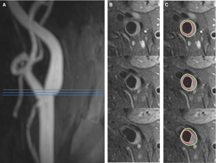
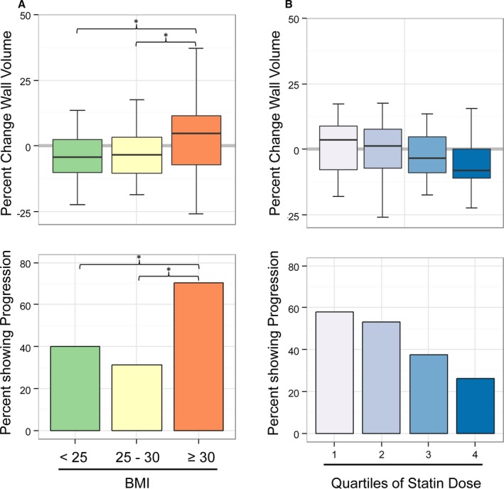
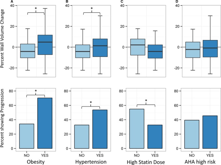
Carotid magnetic resonance imaging was used to evaluate 106 hyperlipidemic participants at baseline and after 12 months of 3-hydroxy-3-methylglutaryl coenzyme A reductase inhibitor (statin) treatment. Multivariable logistic regression was used to determine factors associated with progression (change in carotid wall volume >0) or regression (change ≤0) of carotid atherosclerosis. Computed tomography coronary calcium scores were obtained at baseline for all participants. The median age was 65 years (interquartile range 60-69 years), and 63% of the participants were male. Body mass index >30, elevated C-reactive protein, and hypertension were associated with increased carotid wall volume (obesity: odds ratio for progression 4.6, 95% CI 1.8-12.4, P<0.01; C-reactive protein: odds ratio for progression 2.56, 95% CI 1.17-5.73, P=0.02; hypertension: odds ratio 2.4, 95% CI 1.1-5.3, P<0.05). Higher statin dose was associated with regression of carotid wall volume (P<0.05). In multivariable analysis, obesity remained associated with progression (P<0.01), whereas statin use remained associated with regression (P<0.05). Change in atheroma volume in obese participants was +4.8% versus -4.2% in nonobese participants (P<0.05) despite greater low-density lipoprotein cholesterol reduction in obese participants.
In a population with hyperlipidemia, obese patients showed atheroma progression despite optimized statin therapy.
URL: http://www.clinicaltrials.gov. Unique identifier: NCT01212900.
本研究旨在确定他汀类药物治疗及心血管危险因素与颈动脉粥样硬化变化之间的关系。
采用颈动脉磁共振成像对106名高脂血症参与者在基线时以及接受3-羟基-3-甲基戊二酰辅酶A还原酶抑制剂(他汀类药物)治疗12个月后进行评估。使用多变量逻辑回归来确定与颈动脉粥样硬化进展(颈动脉壁体积变化>0)或消退(变化≤0)相关的因素。所有参与者在基线时均获得了计算机断层扫描冠状动脉钙化评分。中位年龄为65岁(四分位间距60 - 69岁),63%的参与者为男性。体重指数>30、C反应蛋白升高和高血压与颈动脉壁体积增加相关(肥胖:进展的优势比为4.6,95%可信区间1.8 - 12.4,P<0.01;C反应蛋白:进展的优势比为2.56,95%可信区间1.17 - 5.73,P = 0.02;高血压:优势比2.4,95%可信区间1.1 - 5.3,P<0.05)。较高的他汀类药物剂量与颈动脉壁体积消退相关(P<0.05)。在多变量分析中,肥胖仍然与进展相关(P<0.01),而使用他汀类药物仍然与消退相关(P<0.05)。尽管肥胖参与者的低密度脂蛋白胆固醇降低幅度更大,但肥胖参与者的动脉粥样硬化体积变化为+4.8%,而非肥胖参与者为-4.2%(P<0.05)。
在高脂血症人群中,肥胖患者尽管接受了优化的他汀类药物治疗,但仍出现动脉粥样硬化进展。